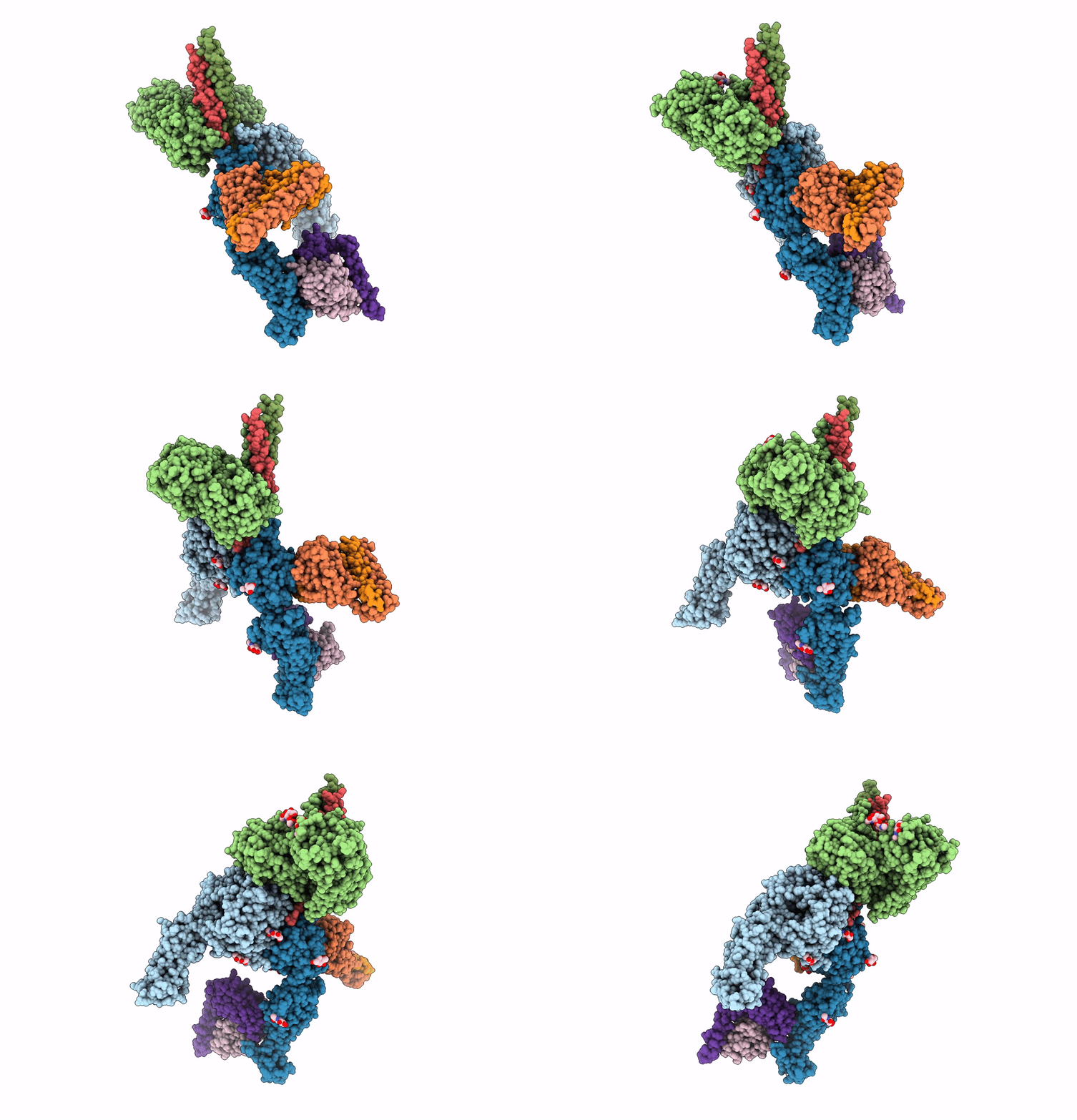
7CEC image
Deposition Date 2020-06-22
Release Date 2021-06-23
Last Version Date 2024-10-16
Entry Detail
PDB ID:
7CEC
Title:
Structure of alpha6beta1 integrin in complex with laminin-511
Biological Source:
Source Organism:
Homo sapiens (Taxon ID: 9606)
Mus musculus (Taxon ID: 10090)
Host Organism:
Method Details:
Experimental Method:
Resolution:
3.90 Å
Aggregation State:
PARTICLE
Reconstruction Method:
SINGLE PARTICLE
Macromolecular Entities
Structures with similar UniProt ID
Protein Blast
Polymer Type:polypeptide(L)
Molecule:Integrin alpha-6
Gene (Uniprot):ITGA6
Chain IDs:A
Chain Length:627
Number of Molecules:1
Biological Source:Homo sapiens
Structures with similar UniProt ID
Protein Blast
Polymer Type:polypeptide(L)
Molecule:Integrin beta-1
Gene (Uniprot):ITGB1
Chain IDs:B
Chain Length:454
Number of Molecules:1
Biological Source:Homo sapiens
Structures with similar UniProt ID
Protein Blast
Polymer Type:polypeptide(L)
Molecule:Laminin subunit alpha-5
Gene (Uniprot):LAMA5
Mutagens:I2723C
Chain IDs:C
Chain Length:674
Number of Molecules:1
Biological Source:Homo sapiens
Structures with similar UniProt ID
Protein Blast
Polymer Type:polypeptide(L)
Molecule:Laminin subunit beta-1
Gene (Uniprot):LAMB1
Chain IDs:D
Chain Length:74
Number of Molecules:1
Biological Source:Homo sapiens
Structures with similar UniProt ID
Protein Blast
Polymer Type:polypeptide(L)
Molecule:Laminin subunit gamma-1
Gene (Uniprot):LAMC1
Mutagens:D1585C
Chain IDs:E
Chain Length:83
Number of Molecules:1
Biological Source:Homo sapiens
Structures with similar UniProt ID
Protein Blast
Polymer Type:polypeptide(L)
Molecule:TS2/16 VH(S112C)-SARAH,TS2/16 VH(S112C)-SARAH
Chain IDs:F
Chain Length:172
Number of Molecules:1
Biological Source:Mus musculus, Homo sapiens
Structures with similar UniProt ID
Protein Blast
Polymer Type:polypeptide(L)
Molecule:TS2/16 VL-SARAH(S37C),TS2/16 VL-SARAH(S37C)
Chain IDs:G
Chain Length:164
Number of Molecules:1
Biological Source:Mus musculus, Homo sapiens
Structures with similar UniProt ID
Protein Blast
Polymer Type:polypeptide(L)
Molecule:HUTS-4 VH(S112C)-SARAH,HUTS-4 VH(S112C)-SARAH
Chain IDs:H
Chain Length:174
Number of Molecules:1
Biological Source:Mus musculus, Homo sapiens
Structures with similar UniProt ID
Protein Blast
Polymer Type:polypeptide(L)
Molecule:HUTS-4 VL(C87Y)-SARAH(S37C),HUTS-4 VL(C87Y)-SARAH(S37C)
Chain IDs:I
Chain Length:159
Number of Molecules:1
Biological Source:Mus musculus, Homo sapiens
Primary Citation
Structural mechanism of laminin recognition by integrin.
Nat Commun 12 4012 4012 (2021)
PMID: 34188035 DOI: 10.1038/s41467-021-24184-8

Abstact

Recognition of laminin by integrin receptors is central to the epithelial cell adhesion to basement membrane, but the structural background of this molecular interaction remained elusive. Here, we report the structures of the prototypic laminin receptor α6β1 integrin alone and in complex with three-chain laminin-511 fragment determined via crystallography and cryo-electron microscopy, respectively. The laminin-integrin interface is made up of several binding sites located on all five subunits, with the laminin γ1 chain C-terminal portion providing focal interaction using two carboxylate anchor points to bridge metal-ion dependent adhesion site of integrin β1 subunit and Asn189 of integrin α6 subunit. Laminin α5 chain also contributes to the affinity and specificity by making electrostatic interactions with large surface on the β-propeller domain of α6, part of which comprises an alternatively spliced X1 region. The propeller sheet corresponding to this region shows unusually high mobility, suggesting its unique role in ligand capture.

Legend

Protein

Chemical

Disease

Primary Citation of related structures