Deposition Date
2020-06-22
Release Date
2021-06-23
Last Version Date
2024-10-30
Entry Detail
PDB ID:
7CEB
Keywords:
Title:
Crystal structure of alpha6beta1 integrin headpiece
Biological Source:
Source Organism(s):
Homo sapiens (Taxon ID: 9606)
Mus musculus (Taxon ID: 10090)
Mus musculus (Taxon ID: 10090)
Expression System(s):
Method Details:
Experimental Method:
Resolution:
2.89 Å
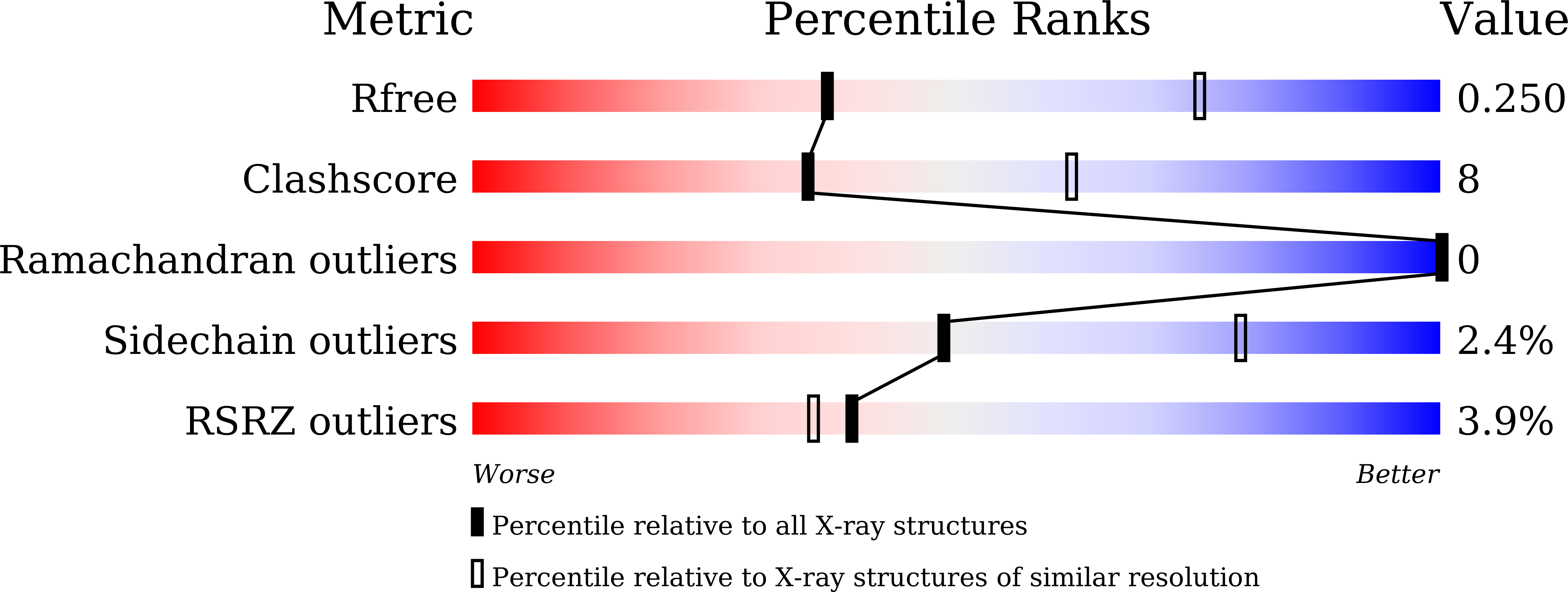
R-Value Free:
0.24
R-Value Work:
0.20
R-Value Observed:
0.20
Space Group:
P 21 21 2