7CD2 image
Deposition Date 2020-06-18
Release Date 2021-03-03
Last Version Date 2024-11-20
Entry Detail
PDB ID:
7CD2
Title:
Crystal structure of the S103F mutant of Bacillus subtilis (natto) YabJ protein.
Biological Source:
Method Details:
Experimental Method:
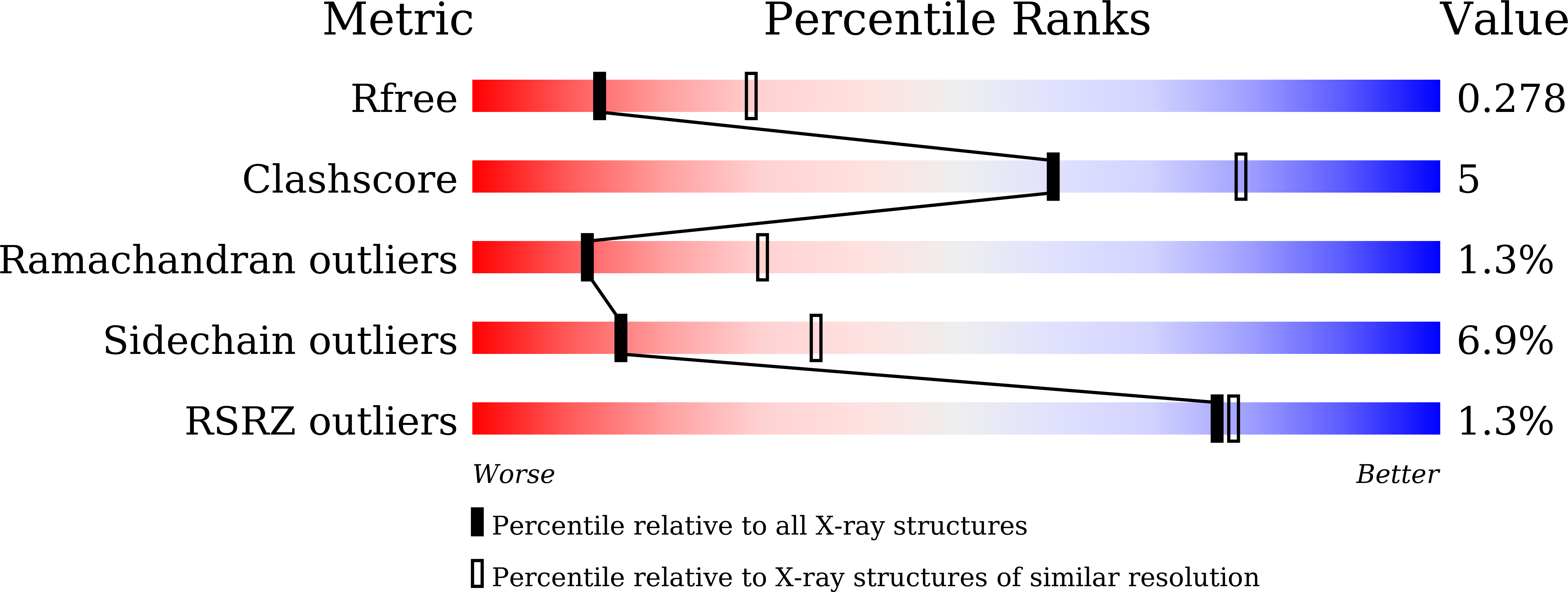
Resolution:
2.70 Å
R-Value Free:
0.27
R-Value Work:
0.18
R-Value Observed:
0.19
Space Group:
P 21 21 21
Macromolecular Entities
Polymer Type:polypeptide(L)
Molecule:YabJ protein
Mutagens:S103F
Chain IDs:A, B, C, D, E, F, G, H, I, J, K, L, M, N, O, P, Q, R, S, T
Chain Length:125
Number of Molecules:20
Biological Source:Bacillus subtilis subsp. natto (strain BEST195)
Ligand Molecules
Primary Citation
Tetramer formation of Bacillus subtilis YabJ protein that belongs to YjgF/YER057c/UK114 family.
Biosci.Biotechnol.Biochem. 85 297 306 (2021)
PMID: 33590041 DOI: 10.1093/bbb/zbaa037

Abstact

Bacillus subtilis YabJ protein belongs to the highly conserved YjgF/YER057c/UK114 family, which has a homotrimeric quaternary structure. The dominant allele of yabJ gene that is caused by a single amino acid mutation of Ser103Phe enables poly-γ-glutamic acid (γPGA) production of B. subtilis under conditions where the cell-density signal transduction was disturbed by the loss of DegQ function. X-ray crystallography of recombinant proteins revealed that unlike the homotrimeric wild-type YabJ, the mutant YabJ(Ser103Phe) had a homotetrameric quaternary structure, and the structural change appeared to be triggered by an inversion of the fifth β-strand. The YabJ homotetramer has a hole that is highly accessible, penetrating through the tetramer, and 2 surface concaves as potential ligand-binding sites. Western blot analyses revealed that the conformational change was also induced in vivo by the Ser103Phe mutation.

Legend

Protein

Chemical

Disease

Primary Citation of related structures
Feedback Form
Name
Email
Institute
Feedback