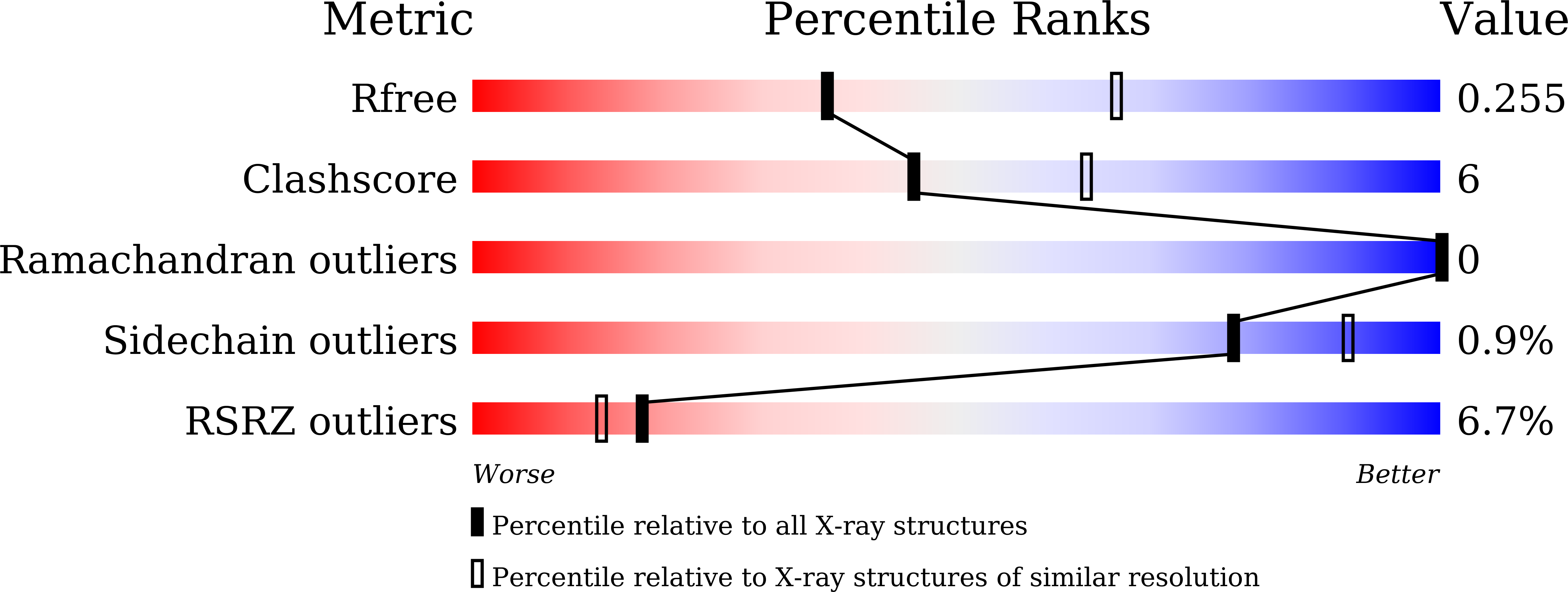
7CBZ image
Deposition Date 2020-06-15
Release Date 2021-06-23
Last Version Date 2023-11-29
Entry Detail
PDB ID:
7CBZ
Title:
Crystal structure of T2R-TTL-A31 complex
Biological Source:
Source Organism:
Sus scrofa (Taxon ID: 9823)
Rattus norvegicus (Taxon ID: 10116)
Gallus gallus (Taxon ID: 9031)
Host Organism:
Method Details:
Experimental Method:
Resolution:
2.61 Å
R-Value Free:
0.25
R-Value Work:
0.20
R-Value Observed:
0.20
Space Group:
P 21 21 21
Macromolecular Entities
Polymer Type:polypeptide(L)
Molecule:Tubulin alpha-1B chain
Gene (Uniprot):TUBA1B
Chain IDs:A, C
Chain Length:451
Number of Molecules:2
Biological Source:Sus scrofa
Polymer Type:polypeptide(L)
Molecule:Tubulin beta chain
Chain IDs:B, D
Chain Length:445
Number of Molecules:2
Biological Source:Sus scrofa
Polymer Type:polypeptide(L)
Molecule:Stathmin-4
Gene (Uniprot):Stmn4
Chain IDs:E
Chain Length:189
Number of Molecules:1
Biological Source:Rattus norvegicus
Polymer Type:polypeptide(L)
Molecule:Tubulin tyrosine ligase
Chain IDs:F
Chain Length:383
Number of Molecules:1
Biological Source:Gallus gallus
Primary Citation
Design, Synthesis, and Bioactivity Evaluation of Dual-Target Inhibitors of Tubulin and Src Kinase Guided by Crystal Structure.
J.Med.Chem. 64 8127 8141 (2021)
PMID: 34081857 DOI: 10.1021/acs.jmedchem.0c01961

Abstact

Klisyri (KX01) is a dual tubulin/Src protein inhibitor that has shown potential therapeutic effects in several tumor models. However, a phase II clinical trial in patients with bone-metastatic castration-resistant prostate cancer was halted because of lack of efficacy. We previously reported that KX01 binds to the colchicine site of β-tubulin and its morpholine group lies close to α-tubulin's surface. Thus, we hypothesized that enhancing the interaction of KX01 with α-tubulin could increase tubulin inhibition and synthesized a series of KX01 derivatives directed by docking studies. Among these derivatives, 8a exhibited more than 10-fold antiproliferation activity in several tumor cells than KX01 and significantly improved in vivo antitumor effects. The X-ray crystal structure suggested that 8a both bound to the colchicine site and extended into the interior of α-tubulin to form potent interactions, presenting a novel binding mode. A potential clinical candidate for cancer therapy was identified in this study.

Legend

Protein

Chemical

Disease

Primary Citation of related structures