7C9B image
Deposition Date 2020-06-05
Release Date 2021-06-09
Last Version Date 2023-11-29
Entry Detail
PDB ID:
7C9B
Keywords:
Title:
Crystal structure of dipeptidase-E from Xenopus laevis
Biological Source:
Source Organism:
Xenopus laevis (Taxon ID: 8355)
Method Details:
Experimental Method:
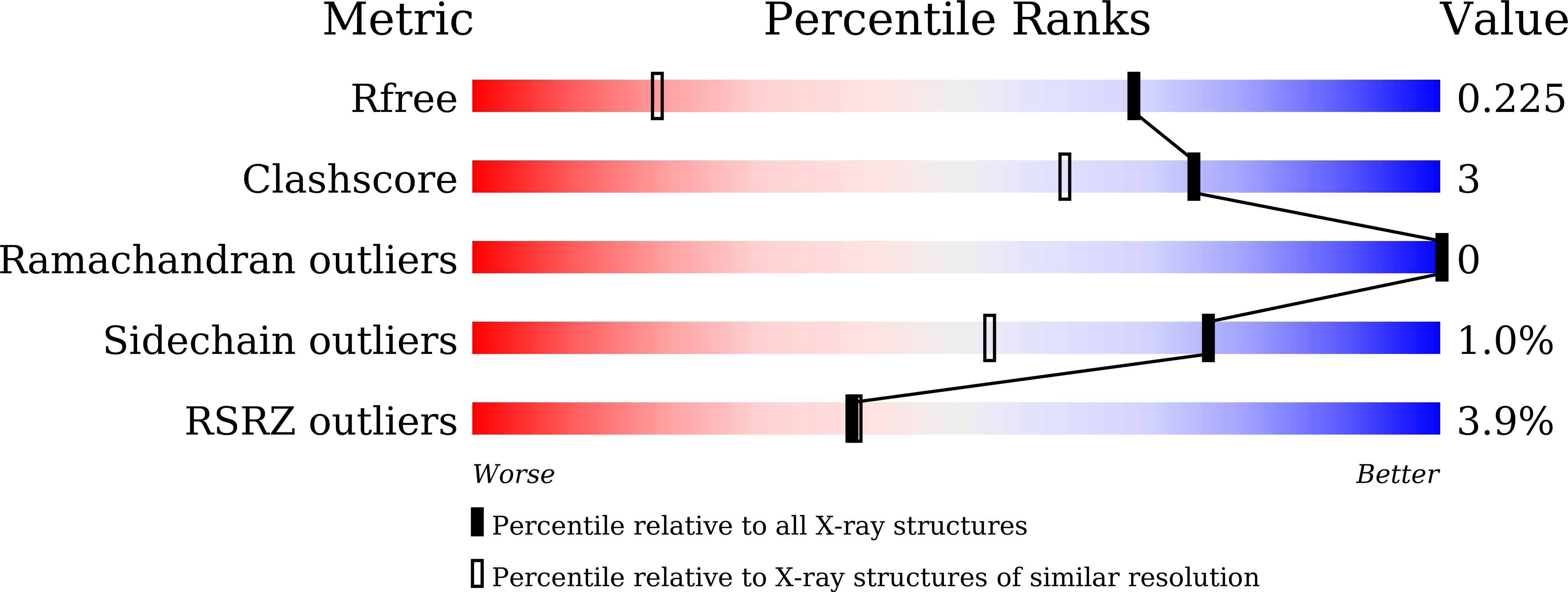
Resolution:
1.40 Å
R-Value Free:
0.22
R-Value Work:
0.19
R-Value Observed:
0.19
Space Group:
C 1 2 1
Macromolecular Entities
Polymer Type:polypeptide(L)
Molecule:Alpha-aspartyl dipeptidase
Gene (Uniprot):aad-a
Chain IDs:A
Chain Length:242
Number of Molecules:1
Biological Source:Xenopus laevis
Primary Citation
Crystal structure of aspartyl dipeptidase from Xenopus laevis revealed ligand binding induced loop ordering and catalytic triad assembly.
Proteins 90 299 308 (2022)
PMID: 34431561 DOI: 10.1002/prot.26220

Abstact

Gene encoding aspartyl dipeptidase from Xenopus levies (PepExl) is upregulated by thyroid hormone and is proposed to play a significant role in resorption of tadpole tail during metamorphosis. However, the importance of peptidase activity for the resorption of the tail remain elusive. Here we report the crystal structures of first eukaryotic S51 peptidase, PepExl, in its ligand-free and Asp-bound states at 1.4 and 1.8 Å resolutions, respectively. The active site is located at dimeric interface and the catalytic triad is found to be dissembled in ligand-free and assembled in Asp-bound state. Structural comparison and molecular dynamic simulations of ligand-free and Asp-bound states shows that distinct loop (loop-A) plays an important role in active site shielding, substrate binding and enzyme activation. This study illuminates the Asp-X dipeptide binding in PepExl is associated with ordering of the loop-A and assembly of residues of catalytic triad in active conformation for enzymatic activity.

Legend

Protein

Chemical

Disease

Primary Citation of related structures