7C8I image
Entry Detail
PDB ID:
7C8I
Keywords:
Title:
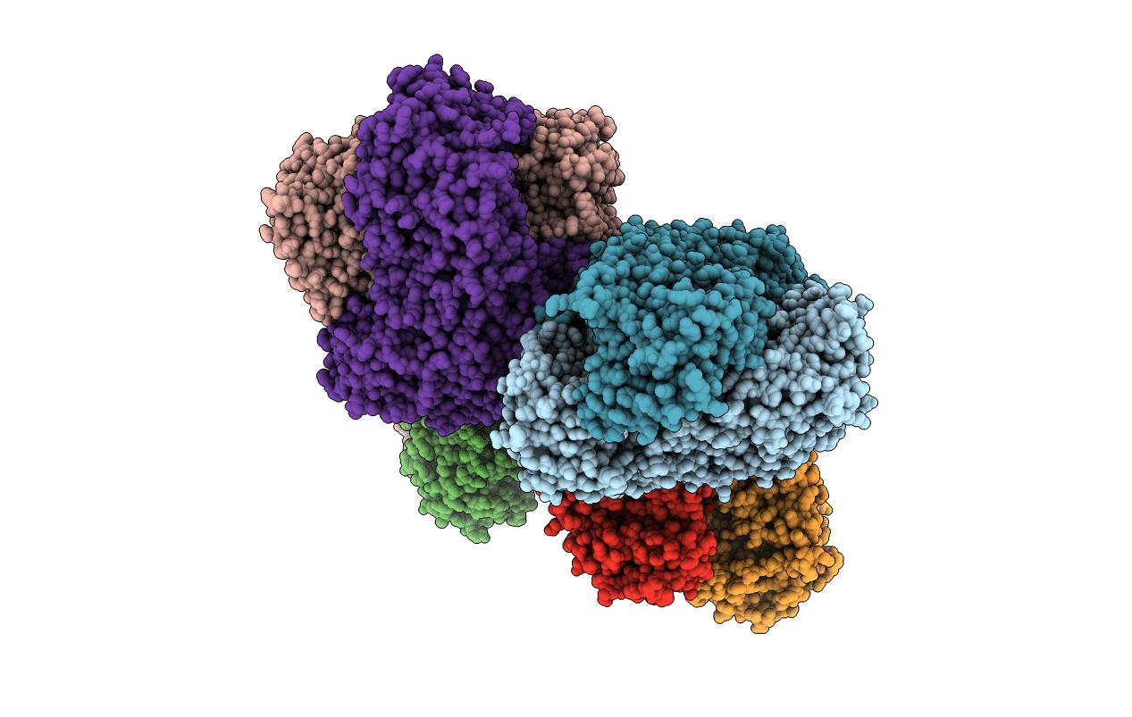
Ambient temperature structure of Bifidobacgterium longum phosphoketolase with thiamine diphosphate and phosphoenol pyuruvate
Biological Source:
Source Organism:
PDB Version:
Deposition Date:
2020-06-01
Release Date:
2021-06-02
Method Details:
Experimental Method:
Resolution:
2.50 Å
R-Value Free:
0.22
R-Value Work:
0.16
R-Value Observed:
0.16
Space Group:
P 1 21 1
Macromolecular Entities
Polymer Type:polypeptide(L)
Description:Xylulose-5-phosphate/fructose-6-phosphate phosphoketolase
Chain IDs:A, B, C, D, E, F, G, H
Chain Length:831
Number of Molecules:8
Biological Source:Bifidobacterium longum
Primary Citation
Ambient temperature structure of phosphoketolase from Bifidobacterium longum determined by serial femtosecond X-ray crystallography.
Acta Crystallogr D Struct Biol 79 290 303 (2023)
PMID: 36974963 DOI: 10.1107/S2059798323001638

Abstact

Phosphoketolase and transketolase are thiamine diphosphate-dependent enzymes and play a central role in the primary metabolism of bifidobacteria: the bifid shunt. The enzymes both catalyze phosphorolytic cleavage of xylulose 5-phosphate or fructose 6-phosphate in the first reaction step, but possess different substrate specificity in the second reaction step, where phosphoketolase and transketolase utilize inorganic phosphate (Pi) and D-ribose 5-phosphate, respectively, as the acceptor substrate. Structures of Bifidobacterium longum phosphoketolase holoenzyme and its complex with a putative inhibitor, phosphoenolpyruvate, were determined at 2.5 Å resolution by serial femtosecond crystallography using an X-ray free-electron laser. In the complex structure, phosphoenolpyruvate was present at the entrance to the active-site pocket and plugged the channel to thiamine diphosphate. The phosphate-group position of phosphoenolpyruvate coincided well with those of xylulose 5-phosphate and fructose 6-phosphate in the structures of their complexes with transketolase. The most striking structural change was observed in a loop consisting of Gln546-Asp547-His548-Asn549 (the QN-loop) at the entrance to the active-site pocket. Contrary to the conformation of the QN-loop that partially covers the entrance to the active-site pocket (`closed form') in the known crystal structures, including the phosphoketolase holoenzyme and its complexes with reaction intermediates, the QN-loop in the current ambient structures showed a more compact conformation with a widened entrance to the active-site pocket (`open form'). In the phosphoketolase reaction, the `open form' QN-loop may play a role in providing the binding site for xylulose 5-phosphate or fructose 6-phosphate in the first step, and the `closed form' QN-loop may help confer specificity for Pi in the second step.

Legend

Protein

Chemical

Disease

Primary Citation of related structures