7C6O image
Deposition Date 2020-05-22
Release Date 2020-12-02
Last Version Date 2023-11-29
Entry Detail
PDB ID:
7C6O
Keywords:
Title:
Catalytic Subunit of Cobaltochelatase from Mycobacterium tuberculosis
Biological Source:
Source Organism:
Host Organism:
Method Details:
Experimental Method:
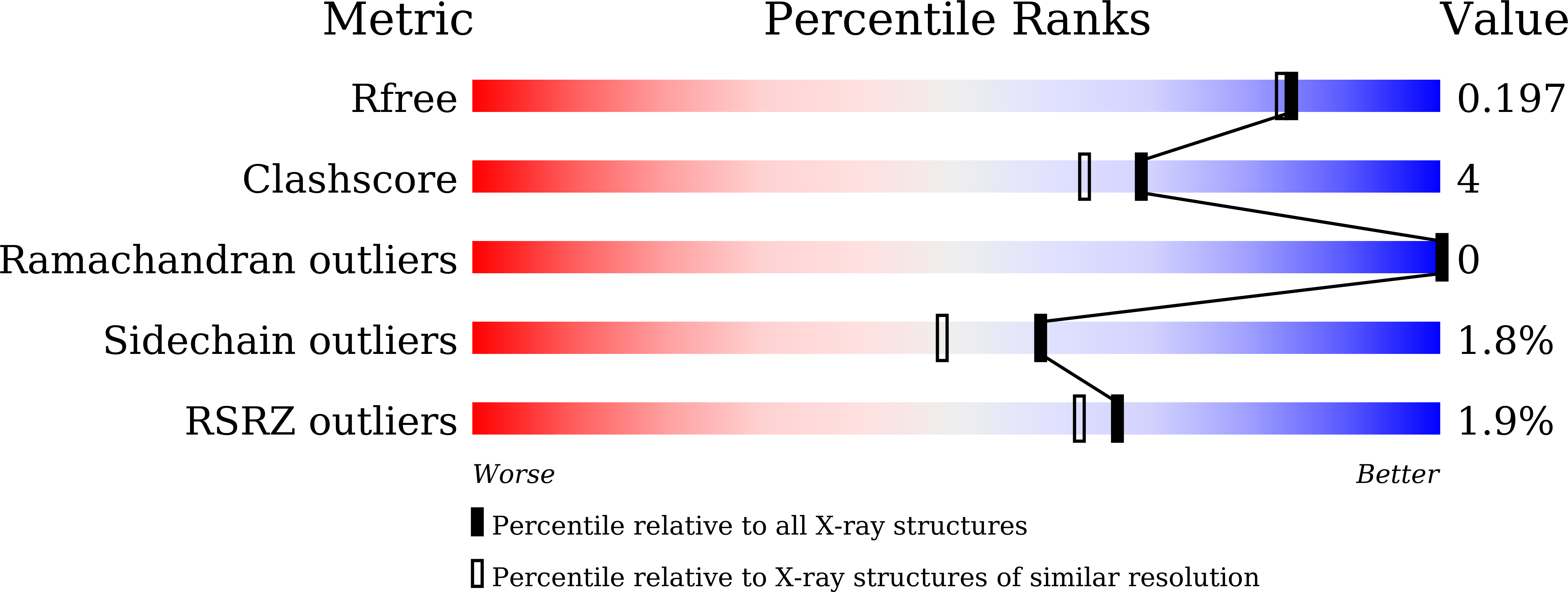
Resolution:
1.80 Å
R-Value Free:
0.19
R-Value Work:
0.16
R-Value Observed:
0.16
Space Group:
P 1 21 1
Macromolecular Entities
Polymer Type:polypeptide(L)
Molecule:Cobalamin biosynthesis protein CobN
Chain IDs:A
Chain Length:1230
Number of Molecules:1
Biological Source:Mycobacterium tuberculosis
Primary Citation
Crystal structure of the large subunit of cobaltochelatase from Mycobacterium tuberculosis.
Proteins 89 462 467 (2021)
PMID: 33210347 DOI: 10.1002/prot.26023

Abstact

Cobaltochelatase in aerobic cobalamin biosynthesis is a complex composed of three subunits. The large subunit CobN is a 140-kDa protein and is homologous to the ChlH subunit of magnesium chelatase. Previously we have reported the 2.5-Å structure of a cyanobacterial ChlH. Here we present the 1.8-Å structure of CobN from Mycobacterium tuberculosis. The overall structure of CobN and ChlH is similar, but significant difference occurs in the head domain. Structural comparison of domains between the two proteins unravels candidate regions for substrate binding and helps to locate a triad of residues that may be essential for metal ion binding.

Legend

Protein

Chemical

Disease

Primary Citation of related structures