7BYX image
Deposition Date 2020-04-24
Release Date 2020-11-04
Last Version Date 2023-11-29
Entry Detail
PDB ID:
7BYX
Keywords:
Title:
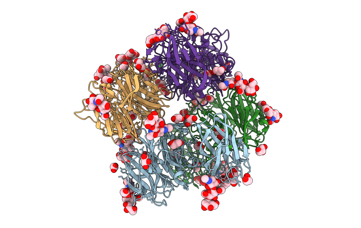
Crystal structure of exo-beta-1,3-galactanase from Phanerochaete chrysosporium Pc1,3Gal43A E208A with beta-1,3-galactotriose
Biological Source:
Source Organism:
Host Organism:
Method Details:
Experimental Method:
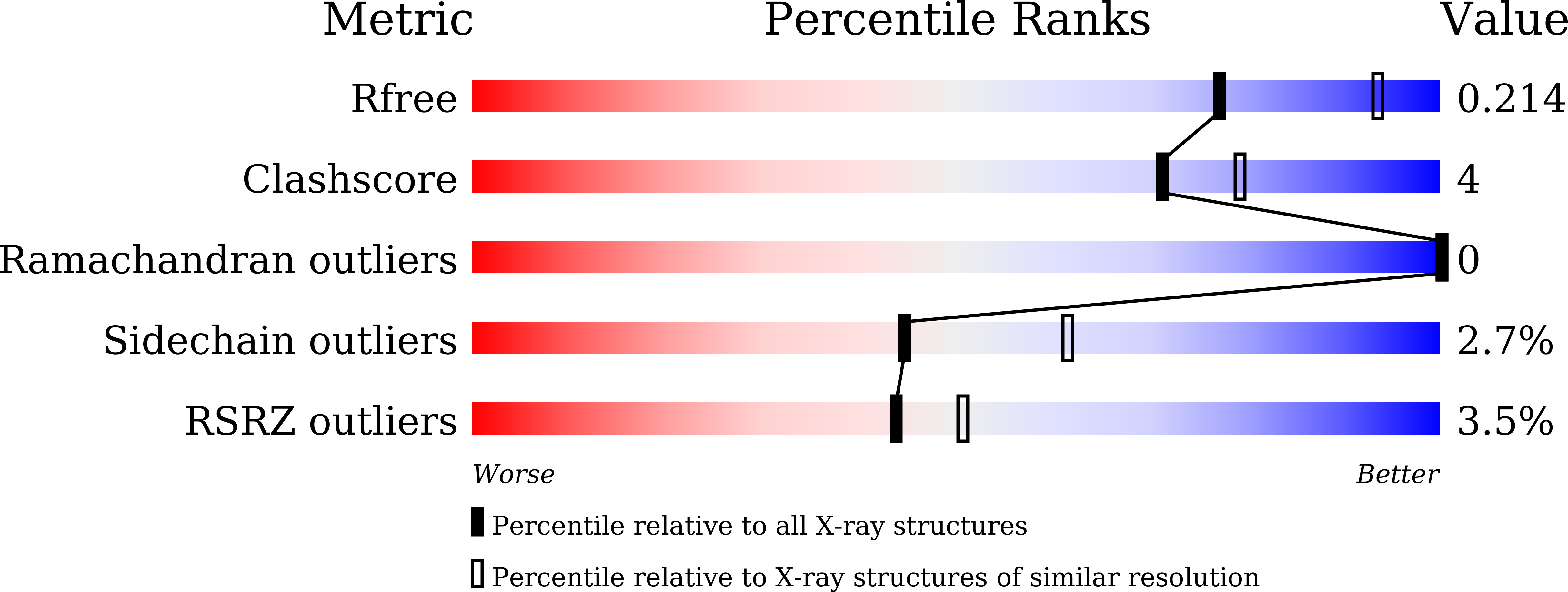
Resolution:
2.30 Å
R-Value Free:
0.21
R-Value Work:
0.16
R-Value Observed:
0.16
Space Group:
P 32 2 1
Macromolecular Entities
Polymer Type:polypeptide(L)
Molecule:Galactan 1,3-beta-galactosidase
Mutations:E208A
Chain IDs:A, B, C, D
Chain Length:427
Number of Molecules:4
Biological Source:Phanerochaete chrysosporium
Primary Citation
Unique active-site and subsite features in the arabinogalactan-degrading GH43 exo-beta-1,3-galactanase from Phanerochaete chrysosporium .
J.Biol.Chem. 295 18539 18552 (2020)
PMID: 33093171 DOI: 10.1074/jbc.RA120.016149

Abstact

Arabinogalactan proteins (AGPs) are plant proteoglycans with functions in growth and development. However, these functions are largely unexplored, mainly because of the complexity of the sugar moieties. These carbohydrate sequences are generally analyzed with the aid of glycoside hydrolases. The exo-β-1,3-galactanase is a glycoside hydrolase from the basidiomycete Phanerochaete chrysosporium (Pc1,3Gal43A), which specifically cleaves AGPs. However, its structure is not known in relation to its mechanism bypassing side chains. In this study, we solved the apo and liganded structures of Pc1,3Gal43A, which reveal a glycoside hydrolase family 43 subfamily 24 (GH43_sub24) catalytic domain together with a carbohydrate-binding module family 35 (CBM35) binding domain. GH43_sub24 is known to lack the catalytic base Asp conserved among other GH43 subfamilies. Our structure in combination with kinetic analyses reveals that the tautomerized imidic acid group of Gln263 serves as the catalytic base residue instead. Pc1,3Gal43A has three subsites that continue from the bottom of the catalytic pocket to the solvent. Subsite -1 contains a space that can accommodate the C-6 methylol of Gal, enabling the enzyme to bypass the β-1,6-linked galactan side chains of AGPs. Furthermore, the galactan-binding domain in CBM35 has a different ligand interaction mechanism from other sugar-binding CBM35s, including those that bind galactomannan. Specifically, we noted a Gly → Trp substitution, which affects pyranose stacking, and an Asp → Asn substitution in the binding pocket, which recognizes β-linked rather than α-linked Gal residues. These findings should facilitate further structural analysis of AGPs and may also be helpful in engineering designer enzymes for efficient biomass utilization.

Legend

Protein

Chemical

Disease

Primary Citation of related structures