7BWN image
Deposition Date 2020-04-15
Release Date 2020-06-17
Last Version Date 2023-11-29
Entry Detail
PDB ID:
7BWN
Keywords:
Title:
Crystal Structure of a Designed Protein Heterocatenane
Biological Source:
Source Organism:
Aequorea victoria (Taxon ID: 6100)
Homo sapiens (Taxon ID: 9606)
Host Organism:
Method Details:
Experimental Method:
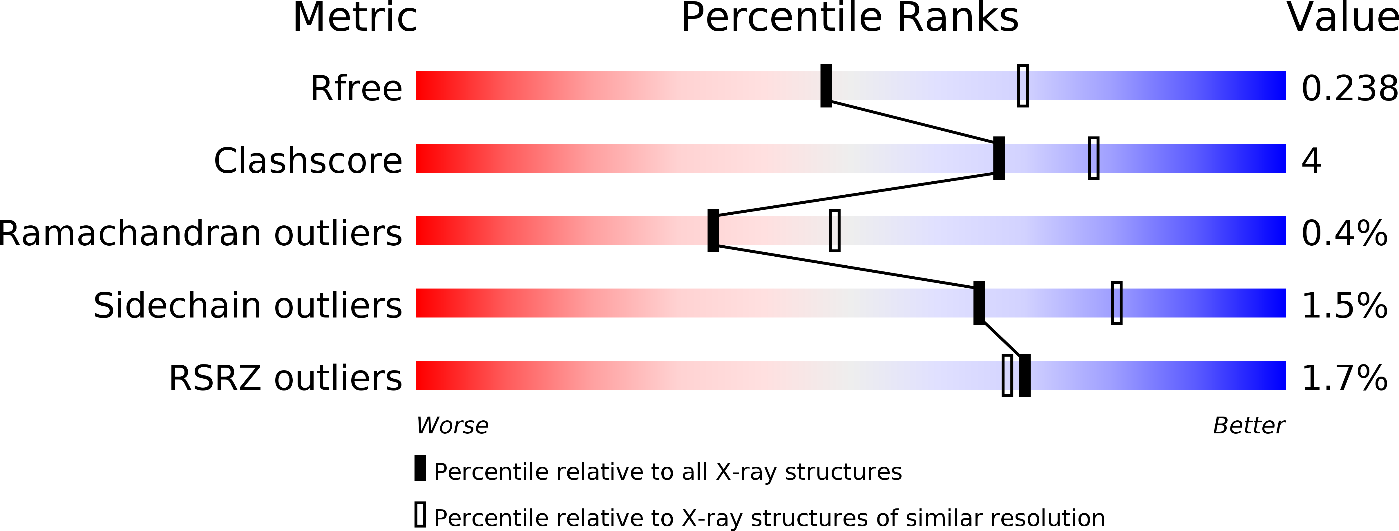
Resolution:
2.40 Å
R-Value Free:
0.23
R-Value Work:
0.18
R-Value Observed:
0.18
Space Group:
H 3
Macromolecular Entities
Protein Blast
Polymer Type:polypeptide(L)
Molecule:Chimera of Green fluorescent protein and p53dim
Mutagens:R2S,S30R,A72S,Q80R,A206V,M340E,L344K
Chain IDs:A (auth: F), C (auth: A), E (auth: C), G (auth: E), I (auth: H), K (auth: J), M, O
Chain Length:280
Number of Molecules:8
Biological Source:Aequorea victoria
Structures with similar UniProt ID
Protein Blast
Polymer Type:polypeptide(L)
Molecule:Cellular tumor antigen p53
Gene (Uniprot):TP53
Chain IDs:B (auth: L), D (auth: B), F (auth: D), H (auth: G), J (auth: I), L (auth: K), N, P
Chain Length:46
Number of Molecules:8
Biological Source:Homo sapiens
Primary Citation
Cellular Synthesis and X-ray Crystal Structure of a Designed Protein Heterocatenane.
Angew.Chem.Int.Ed.Engl. 59 16122 16127 (2020)
PMID: 32506656 DOI: 10.1002/anie.202005490

Abstact

Herein, we report the biosynthesis of protein heterocatenanes using a programmed sequence of multiple post-translational processing events including intramolecular chain entanglement, in situ backbone cleavage, and spontaneous cyclization. The approach is general, autonomous, and can obviate the need for any additional enzymes. The catenane topology was convincingly proven using a combination of SDS-PAGE, LC-MS, size exclusion chromatography, controlled proteolytic digestion, and protein crystallography. The X-ray crystal structure clearly shows two mechanically interlocked protein rings with intact folded domains. It opens new avenues in the nascent field of protein-topology engineering.

Legend

Protein

Chemical

Disease

Primary Citation of related structures