7BVE image
Deposition Date 2020-04-10
Release Date 2020-04-29
Last Version Date 2024-11-06
Entry Detail
PDB ID:
7BVE
Keywords:
Title:
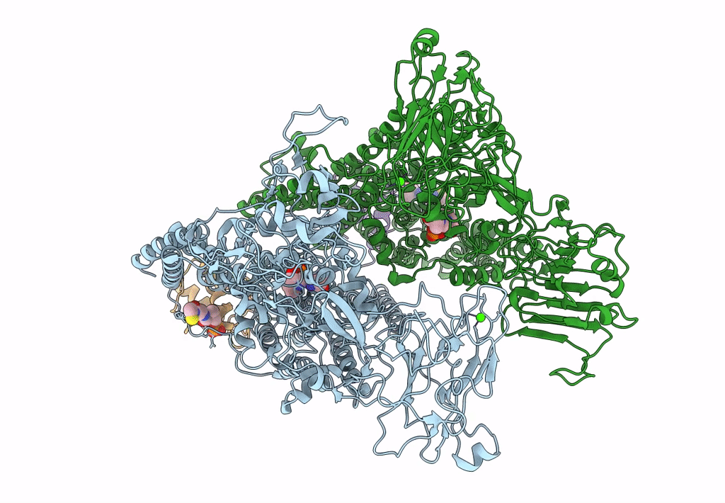
Cryo-EM structure of Mycobacterium smegmatis arabinosyltransferase EmbC2-AcpM2 in complex with ethambutol
Biological Source:
Method Details:
Experimental Method:
Resolution:
2.81 Å
Aggregation State:
PARTICLE
Reconstruction Method:
SINGLE PARTICLE
Macromolecular Entities
Polymer Type:polypeptide(L)
Molecule:Integral membrane indolylacetylinositol arabinosyltransferase EmbC
Chain IDs:A, B
Chain Length:1084
Number of Molecules:2
Biological Source:Mycolicibacterium smegmatis (strain ATCC 700084 / mc(2)155)
Polymer Type:polypeptide(L)
Molecule:Meromycolate extension acyl carrier protein
Gene (Uniprot):acpM
Chain IDs:C, D
Chain Length:99
Number of Molecules:2
Biological Source:Mycolicibacterium smegmatis MC2 155
Primary Citation
Structures of cell wall arabinosyltransferases with the anti-tuberculosis drug ethambutol.
Science 368 1211 1219 (2020)
PMID: 32327601 DOI: 10.1126/science.aba9102

Abstact

The arabinosyltransferases EmbA, EmbB, and EmbC are involved in Mycobacterium tuberculosis cell wall synthesis and are recognized as targets for the anti-tuberculosis drug ethambutol. In this study, we determined cryo-electron microscopy and x-ray crystal structures of mycobacterial EmbA-EmbB and EmbC-EmbC complexes in the presence of their glycosyl donor and acceptor substrates and with ethambutol. These structures show how the donor and acceptor substrates bind in the active site and how ethambutol inhibits arabinosyltransferases by binding to the same site as both substrates in EmbB and EmbC. Most drug-resistant mutations are located near the ethambutol binding site. Collectively, our work provides a structural basis for understanding the biochemical function and inhibition of arabinosyltransferases and the development of new anti-tuberculosis agents.

Legend

Protein

Chemical

Disease

Primary Citation of related structures