7BTK image
Deposition Date 2020-04-01
Release Date 2021-03-03
Last Version Date 2023-11-29
Entry Detail
PDB ID:
7BTK
Keywords:
Title:
E.coli beta-galactosidase (E537Q) in complex with fluorescent probe KSA01
Biological Source:
Source Organism:
Host Organism:
Method Details:
Experimental Method:
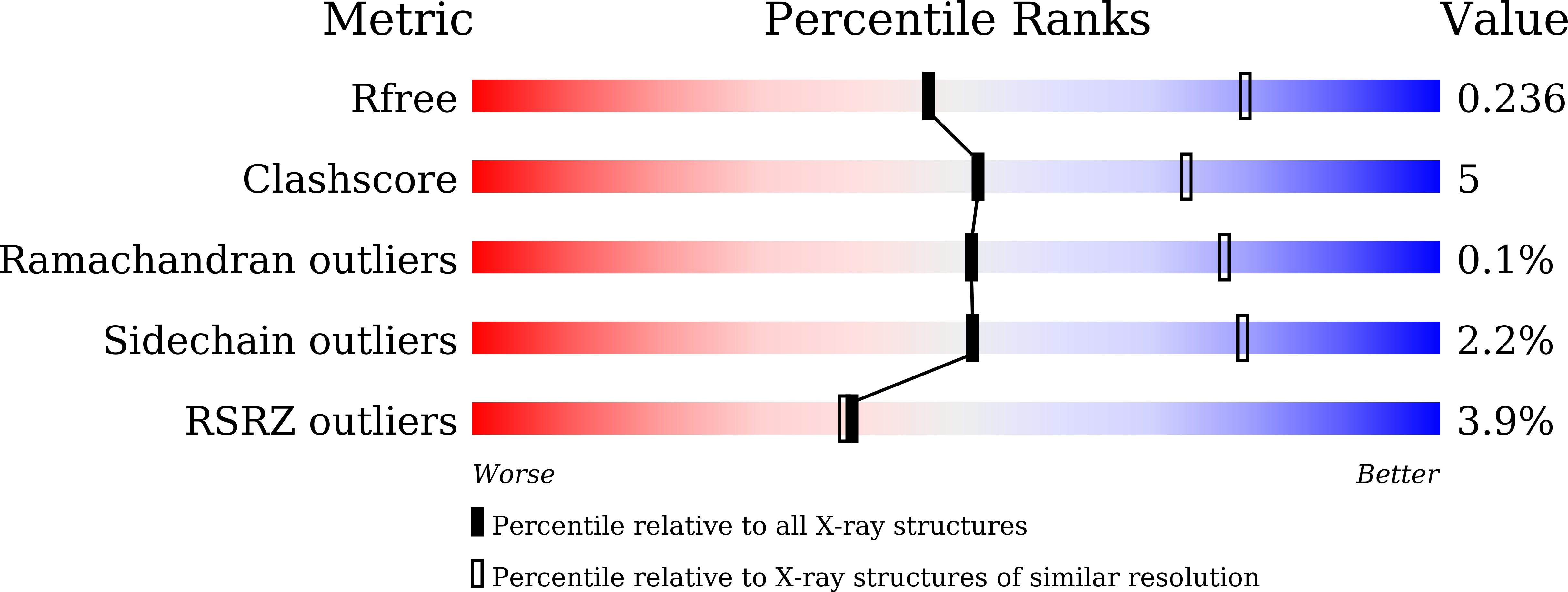
Resolution:
2.70 Å
R-Value Free:
0.23
R-Value Work:
0.21
R-Value Observed:
0.21
Space Group:
C 1 2 1
Macromolecular Entities
Structures with similar UniProt ID
Protein Blast
Polymer Type:polypeptide(L)
Molecule:Beta-galactosidase
Gene (Uniprot):lacZ
Mutagens:E537Q
Chain IDs:A, B, C, D
Chain Length:1025
Number of Molecules:4
Biological Source:Escherichia coli K-12
Primary Citation
Two-Dimensional Design Strategy to Construct Smart Fluorescent Probes for the Precise Tracking of Senescence.
Angew.Chem.Int.Ed.Engl. 60 10756 10765 (2021)
PMID: 33624914 DOI: 10.1002/anie.202101278

Abstact

The tracking of cellular senescence usually depends on the detection of senescence-associated β-galactosidase (SA-β-gal). Previous probes for SA-β-gal with this purpose only cover a single dimension: the accumulation of this enzyme in lysosomes. However, this is insufficient to determine the destiny of senescence because endogenous β-gal enriched in lysosomes is not only related to senescence, but also to some other physiological processes. To address this issue, we introduce our fluorescent probes including a second dimension: lysosomal pH, since de-acidification is a unique feature of the lysosomes in senescent cells. With this novel design, our probes achieved excellent discrimination of SA-β-gal from cancer-associated β-gal, which enables them to track cellular senescence as well as tissue aging more precisely. Our crystal structures of a model enzyme E. coli β-gal mutant (E537Q) complexed with each probe further revealed the structural basis for probe recognition.

Legend

Protein

Chemical

Disease

Primary Citation of related structures