7BRN image
Deposition Date 2020-03-29
Release Date 2020-07-08
Last Version Date 2023-11-29
Entry Detail
PDB ID:
7BRN
Title:
Crystal structure of Atg40 AIM fused to Atg8
Biological Source:
Source Organism:
Method Details:
Experimental Method:
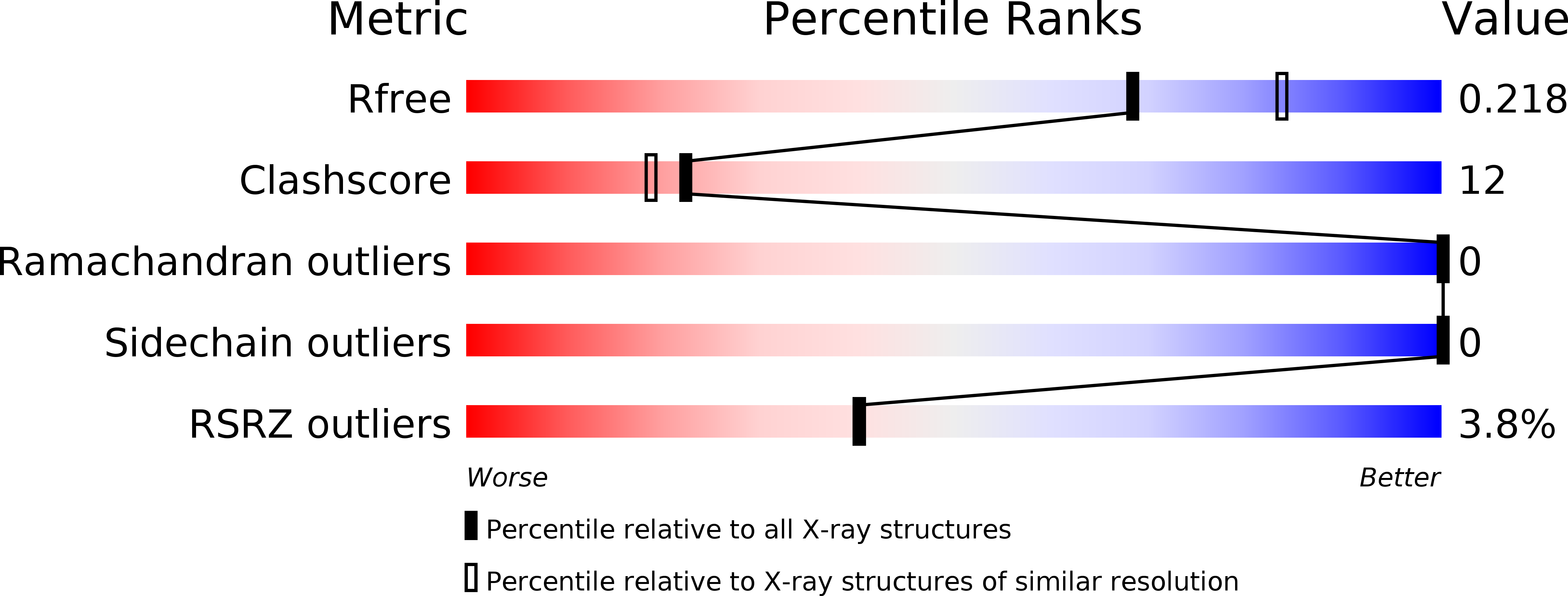
Resolution:
2.23 Å
R-Value Free:
0.21
R-Value Work:
0.20
R-Value Observed:
0.20
Space Group:
P 32 2 1
Macromolecular Entities
Polymer Type:polypeptide(L)
Molecule:Autophagy-related protein 40,Autophagy-related protein 8
Gene (Uniprot):ATG8, ATG40
Mutations:K26P
Chain IDs:A
Chain Length:134
Number of Molecules:1
Biological Source:Saccharomyces cerevisiae S288C
Primary Citation
Super-assembly of ER-phagy receptor Atg40 induces local ER remodeling at contacts with forming autophagosomal membranes.
Nat Commun 11 3306 3306 (2020)
PMID: 32620754 DOI: 10.1038/s41467-020-17163-y

Abstact

The endoplasmic reticulum (ER) is selectively degraded by autophagy (ER-phagy) through proteins called ER-phagy receptors. In Saccharomyces cerevisiae, Atg40 acts as an ER-phagy receptor to sequester ER fragments into autophagosomes by binding Atg8 on forming autophagosomal membranes. During ER-phagy, parts of the ER are morphologically rearranged, fragmented, and loaded into autophagosomes, but the mechanism remains poorly understood. Here we find that Atg40 molecules assemble in the ER membrane concurrently with autophagosome formation via multivalent interaction with Atg8. Atg8-mediated super-assembly of Atg40 generates highly-curved ER regions, depending on its reticulon-like domain, and supports packing of these regions into autophagosomes. Moreover, tight binding of Atg40 to Atg8 is achieved by a short helix C-terminal to the Atg8-family interacting motif, and this feature is also observed for mammalian ER-phagy receptors. Thus, this study significantly advances our understanding of the mechanisms of ER-phagy and also provides insights into organelle fragmentation in selective autophagy of other organelles.

Legend

Protein

Chemical

Disease

Primary Citation of related structures