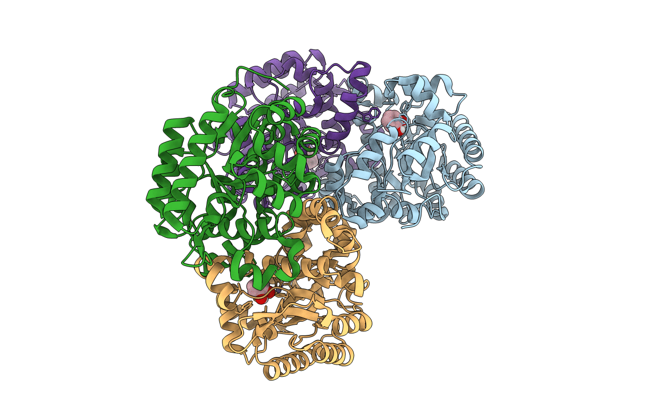
7BP1 image
Entry Detail
PDB ID:
7BP1
Title:
Crystal structure of 2, 3-dihydroxybenzoic acid decarboxylase from Fusarium oxysporum in complex with Catechol
Biological Source:
Source Organism:
Host Organism:
PDB Version:
Deposition Date:
2020-03-21
Release Date:
2020-07-15
Method Details:
Experimental Method:
Resolution:
1.97 Å
R-Value Free:
0.19
R-Value Work:
0.16
R-Value Observed:
0.16
Space Group:
P 21 21 21
Macromolecular Entities
Polymer Type:polypeptide(L)
Description:2,3-dihydroxybenzoate decarboxylase
Chain IDs:A, B, C, D
Chain Length:343
Number of Molecules:4
Biological Source:Fusarium oxysporum
Primary Citation
2,3-Dihydroxybenzoic Acid Decarboxylase from Fusarium oxysporum: Crystal Structures and Substrate Recognition Mechanism.
Chembiochem 21 2950 2956 (2020)
PMID: 32421914 DOI: 10.1002/cbic.202000244

Abstact

A 2,3-dihydroxybenzoic acid decarboxylase from Fusarium oxysporum (2,3-DHBD_Fo) has a relatively high catalytic efficiency for the decarboxylation of 2,3-dihydroxybenzoic acid (DHBA) and carboxylation of catechol, thus it has a different substrate spectrum from other benzoic acid decarboxylases. We have determined the structures of 2,3-DHBD_Fo in its apo form and complexes with catechol or 2,5-dihydroxybenzoic acid at 1.55, 1.97, and 2.45 Å resolution, respectively. The crystal structures of 2,3-DHBD_Fo show that the enzyme exists as a homotetramer, and each active center has a Zn2+ ion coordinated by E8, H167, D291 and three water molecules. This is different from 2,6-DHBD from Rhizobium sporomusa, in which the Zn2+ ion is also coordinated with H10. Surprisingly, mutation of A10 of 2,3-DHBD_Fo to His resulted in almost complete loss of the enzyme activity. Enzyme-substrate docking and site-directed mutation studies indicate that residue R233Δ interacts with the 3-hydroxy group of 2,3-DHBA, and plays an important role in substrate recognition for this enzyme, thus revealing the molecular basis 2,3-dihydroxybenzoic acid decarboxylase.

Legend

Protein

Chemical

Disease

Primary Citation of related structures