7BNY image
Deposition Date 2021-01-22
Release Date 2021-12-08
Last Version Date 2024-11-13
Entry Detail
PDB ID:
7BNY
Keywords:
Title:
Structure of 2A protein from encephalomyocarditis virus (EMCV)
Biological Source:
Source Organism:
Method Details:
Experimental Method:
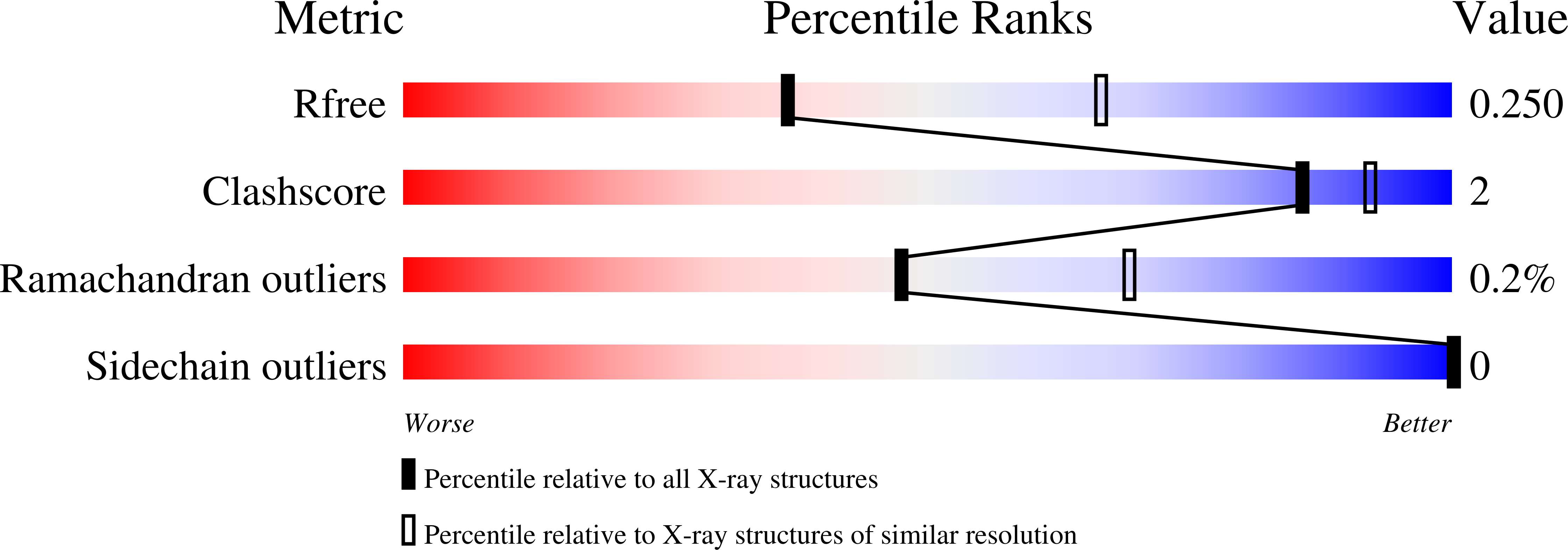
Resolution:
2.62 Å
R-Value Free:
0.25
R-Value Work:
0.22
R-Value Observed:
0.22
Space Group:
P 62 2 2
Macromolecular Entities
Structures with similar UniProt ID
Protein Blast
Polymer Type:polypeptide(L)
Molecule:Genome polyprotein
Chain IDs:A, B, C, D
Chain Length:152
Number of Molecules:4
Biological Source:Mengo encephalomyocarditis virus
Ligand Molecules
Primary Citation
Structural and molecular basis for Cardiovirus 2A protein as a viral gene expression switch.
Nat Commun 12 7166 7166 (2021)
PMID: 34887415 DOI: 10.1038/s41467-021-27400-7

Abstact

Programmed -1 ribosomal frameshifting (PRF) in cardioviruses is activated by the 2A protein, a multi-functional virulence factor that also inhibits cap-dependent translational initiation. Here we present the X-ray crystal structure of 2A and show that it selectively binds to a pseudoknot-like conformation of the PRF stimulatory RNA element in the viral genome. Using optical tweezers, we demonstrate that 2A stabilises this RNA element, likely explaining the increase in PRF efficiency in the presence of 2A. Next, we demonstrate a strong interaction between 2A and the small ribosomal subunit and present a cryo-EM structure of 2A bound to initiated 70S ribosomes. Multiple copies of 2A bind to the 16S rRNA where they may compete for binding with initiation and elongation factors. Together, these results define the structural basis for RNA recognition by 2A, show how 2A-mediated stabilisation of an RNA pseudoknot promotes PRF, and reveal how 2A accumulation may shut down translation during virus infection.

Legend

Protein

Chemical

Disease

Primary Citation of related structures
Feedback Form
Name
Email
Institute
Feedback