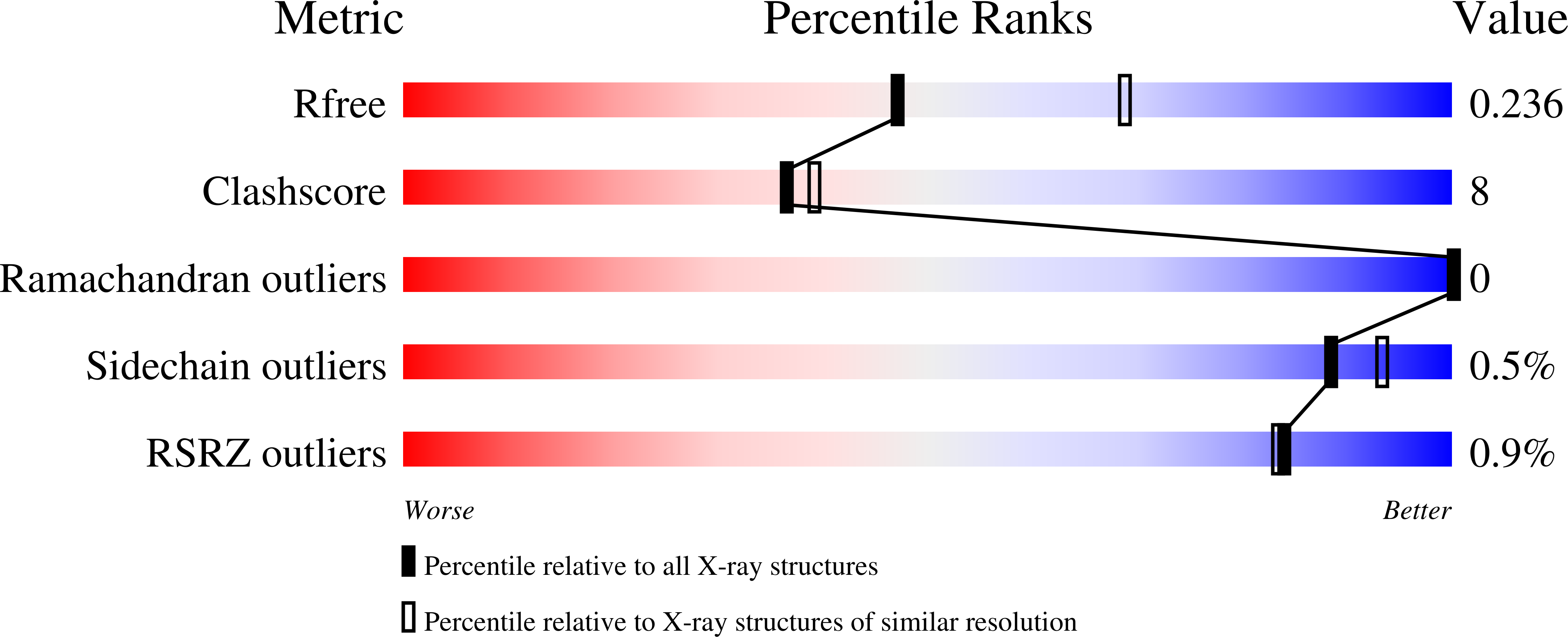
7BGU image
Deposition Date 2021-01-08
Release Date 2021-12-15
Last Version Date 2024-11-13
Entry Detail
PDB ID:
7BGU
Keywords:
Title:
Mason-Pfizer Monkey Virus Protease mutant C7A/D26N/C106A in complex with peptidomimetic inhibitor
Biological Source:
Method Details:
Experimental Method:
Resolution:
2.43 Å
R-Value Free:
0.23
R-Value Work:
0.17
R-Value Observed:
0.18
Space Group:
P 1
Macromolecular Entities
Polymer Type:polypeptide(L)
Molecule:Gag-Pro-Pol polyprotein
Gene (Uniprot):gag-pro-pol
Mutations:C7A, D26N, C106A; ENGINEERED MUTATIONS
Chain IDs:A, B, C, D
Chain Length:114
Number of Molecules:4
Biological Source:Mason-Pfizer monkey virus
Polymer Type:polypeptide(L)
Molecule:peptidomimetic inhibitor
Chain IDs:E (auth: G), F
Chain Length:7
Number of Molecules:2
Biological Source:synthetic construct
Ligand Molecules
Primary Citation

Abstact

Mason-Pfizer monkey virus protease (PR) was crystallized in complex with two pepstatin-based inhibitors in P1 space group. In both crystal structures, the extended flap loops that lock the inhibitor/substrate over the active site, are visible in the electron density either completely or with only small gaps, providing the first observation of the conformation of the flap loops in dimeric complex form of this retropepsin. The H-bond network in the active site (with D26N mutation) differs from that reported for the P21 crystal structures and is similar to a rarely occurring system in HIV-1 PR.

Legend

Protein

Chemical

Disease

Primary Citation of related structures