Deposition Date
2020-12-18
Release Date
2021-10-20
Last Version Date
2024-10-16
Entry Detail
PDB ID:
7BBT
Keywords:
Title:
Structure of cytochrome c in complex with a p-benzyl-sulfonato-calix[8]arene-PEG pseudorotaxane
Biological Source:
Source Organism(s):
Saccharomyces cerevisiae S288C (Taxon ID: 559292)
Expression System(s):
Method Details:
Experimental Method:
Resolution:
3.02 Å
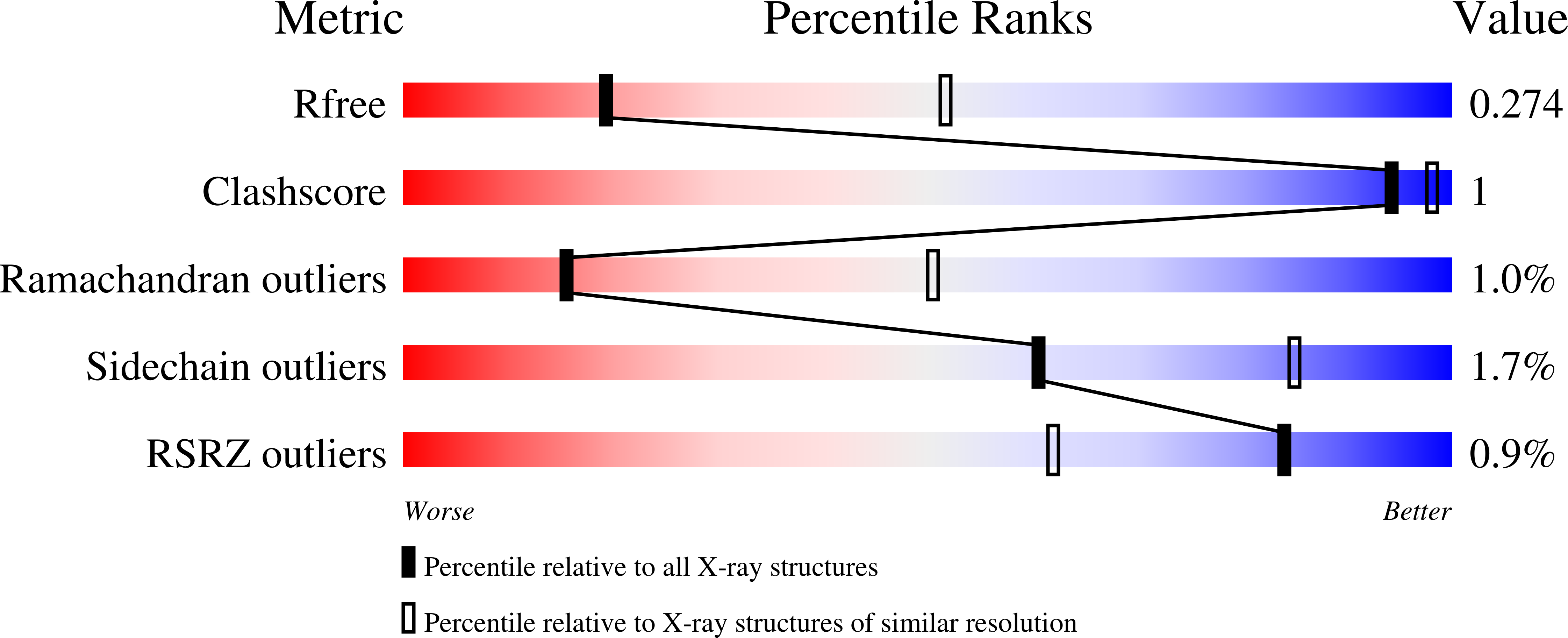
R-Value Free:
0.26
R-Value Work:
0.23
R-Value Observed:
0.23
Space Group:
C 1 2 1