7B9P image
Entry Detail
PDB ID:
7B9P
Keywords:
Title:
Structure of Ribonucleotide reductase from Rhodobacter sphaeroides
Biological Source:
PDB Version:
Deposition Date:
2020-12-14
Release Date:
2022-01-12
Method Details:
Experimental Method:
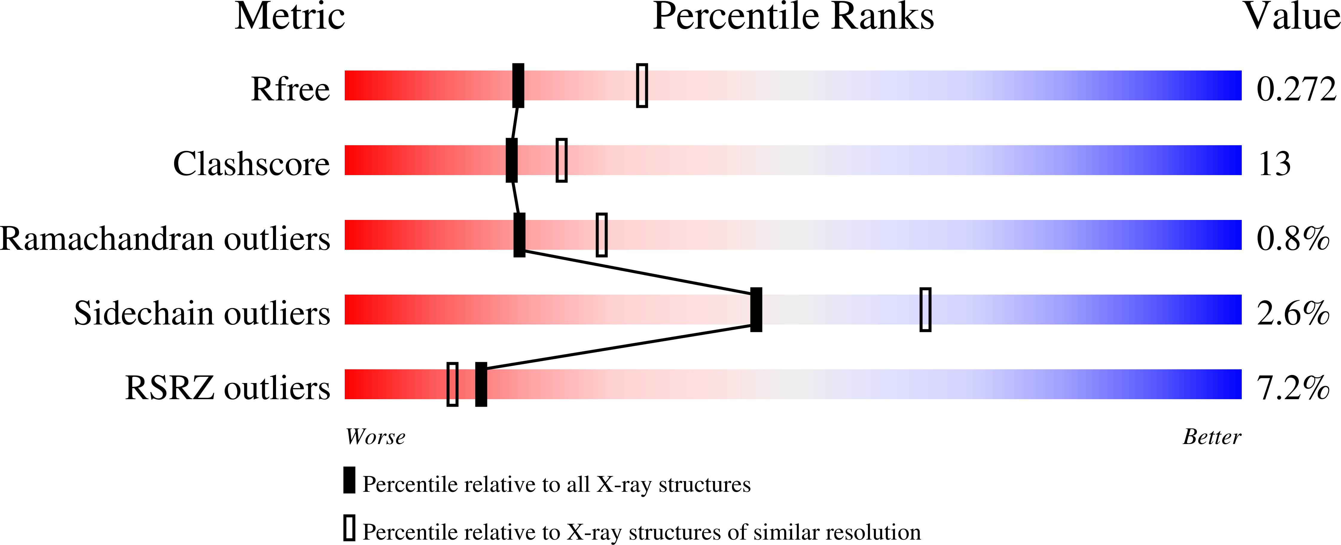
Resolution:
2.65 Å
R-Value Free:
0.27
R-Value Work:
0.22
R-Value Observed:
0.23
Space Group:
P 65 2 2
Macromolecular Entities
Polymer Type:polypeptide(L)
Description:Vitamin B12-dependent ribonucleotide reductase
Mutations:V926Stop
Chain IDs:A
Chain Length:925
Number of Molecules:1
Biological Source:Rhodobacter sphaeroides (strain ATCC 17023 / 2.4.1 / NCIB 8253 / DSM 158)
Primary Citation
HUG Domain Is Responsible for Active Dimer Stabilization in an NrdJd Ribonucleotide Reductase.
Biochemistry 61 1633 1641 (2022)
PMID: 35856337 DOI: 10.1021/acs.biochem.2c00173

Abstact

Ribonucleotide reductases (RNRs) catalyze the reduction of ribonucleotides to the corresponding deoxyribonucleotides. The catalytic activity of most RNRs depends on the formation of a dimer of the catalytic subunits. The active site is located at the interface, and part of the substrate binding site and regulatory mechanisms work across the subunit in the dimer. In this study, we describe and characterize a novel domain responsible for forming the catalytic dimer in several class II RNRs. The 3D structure of the class II RNR from Rhodobacter sphaeroides reveals a so far undescribed α-helical domain in the dimer interface, which is embracing the other subunit. Genetic removal of this HUG domain leads to a severe reduction of activity paired with reduced dimerization capability. In comparison with other described RNRs, the enzyme with this domain is less dependent on the presence of nucleotides to act as allosteric effectors in the formation of dimers. The HUG domain appears to serve as an interlock to keep the dimer intact and functional even at low enzyme and/or effector concentrations.

Legend

Protein

Chemical

Disease

Primary Citation of related structures