Deposition Date
2020-12-07
Release Date
2021-08-11
Last Version Date
2024-07-10
Entry Detail
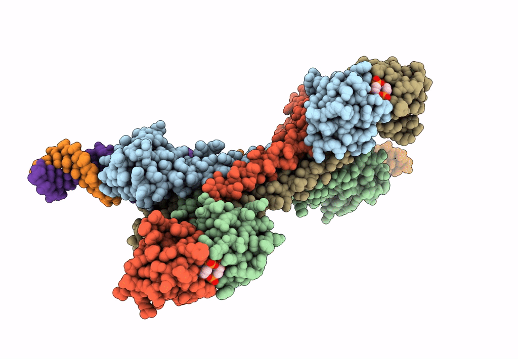
PDB ID:
7B5Y
Keywords:
Title:
S. agalactiae BusR in complex with its busAB-promotor DNA
Biological Source:
Source Organism(s):
Streptococcus agalactiae (Taxon ID: 1311)
Expression System(s):
Method Details:
Experimental Method:
Resolution:
7.10 Å
Aggregation State:
PARTICLE
Reconstruction Method:
SINGLE PARTICLE