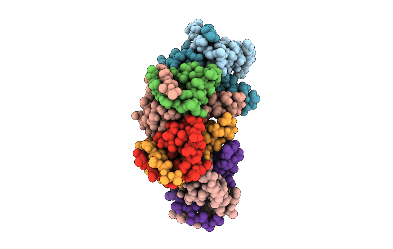
7B22 image
Deposition Date 2020-11-25
Release Date 2021-10-06
Last Version Date 2024-01-31
Entry Detail
PDB ID:
7B22
Title:
Vibrio cholerae ParD2 Antitoxin
Biological Source:
Method Details:
Experimental Method:
Resolution:
3.08 Å
R-Value Free:
0.29
R-Value Work:
0.27
R-Value Observed:
0.27
Space Group:
C 2 2 21
Macromolecular Entities
Structures with similar UniProt ID
Protein Blast
Polymer Type:polypeptide(L)
Molecule:Antitoxin ParD
Gene (Uniprot):parD
Chain IDs:A, B, C, D, E, F, G, H
Chain Length:80
Number of Molecules:8
Biological Source:Vibrio cholerae serotype O1 (strain ATCC 39315 / El Tor Inaba N16961)
Ligand Molecules
Primary Citation
Entropic pressure controls the oligomerization of the Vibrio cholerae ParD2 antitoxin.
Acta Crystallogr D Struct Biol 77 904 920 (2021)
PMID: 34196617 DOI: 10.1107/S2059798321004873

Abstact

ParD2 is the antitoxin component of the parDE2 toxin-antitoxin module from Vibrio cholerae and consists of an ordered DNA-binding domain followed by an intrinsically disordered ParE-neutralizing domain. In the absence of the C-terminal intrinsically disordered protein (IDP) domain, V. cholerae ParD2 (VcParD2) crystallizes as a doughnut-shaped hexadecamer formed by the association of eight dimers. This assembly is stabilized via hydrogen bonds and salt bridges rather than by hydrophobic contacts. In solution, oligomerization of the full-length protein is restricted to a stable, open decamer or dodecamer, which is likely to be a consequence of entropic pressure from the IDP tails. The relative positioning of successive VcParD2 dimers mimics the arrangement of Streptococcus agalactiae CopG dimers on their operator and allows an extended operator to wrap around the VcParD2 oligomer.

Legend

Protein

Chemical

Disease

Primary Citation of related structures