Deposition Date
2020-11-24
Release Date
2021-03-17
Last Version Date
2024-11-13
Entry Detail
PDB ID:
7B1H
Keywords:
Title:
Monoclinic P21 Structure of Human Mad1 C-terminal Domain in Complex with Phosphorylated Bub1 CD1 Domain
Biological Source:
Source Organism(s):
Homo sapiens (Taxon ID: 9606)
Expression System(s):
Method Details:
Experimental Method:
Resolution:
2.40 Å
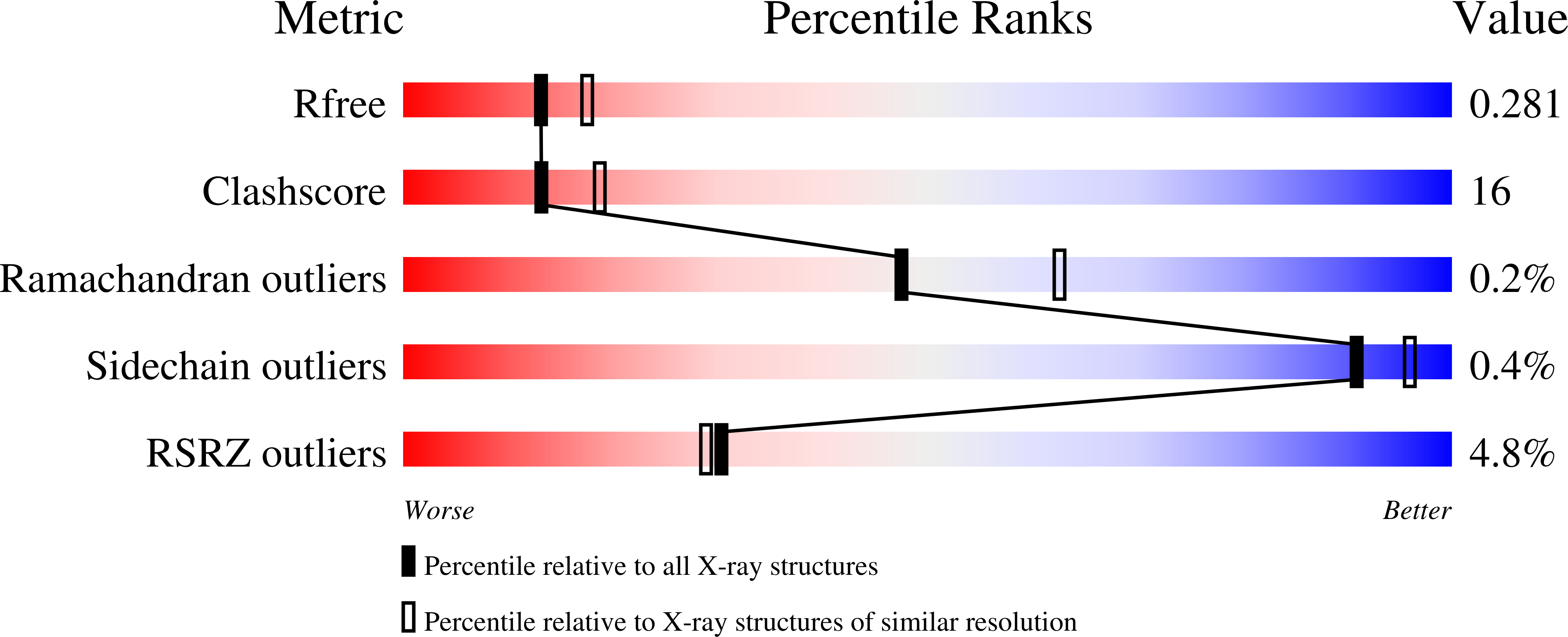
R-Value Free:
0.28
R-Value Work:
0.23
R-Value Observed:
0.23
Space Group:
P 1 21 1