7AYV image
Deposition Date 2020-11-13
Release Date 2021-08-18
Last Version Date 2024-01-31
Entry Detail
PDB ID:
7AYV
Keywords:
Title:
X-ray crystallographic structure of (6-4)photolyase from Drosophila melanogaster at cryogenic temperature
Biological Source:
Source Organism:
Host Organism:
Method Details:
Experimental Method:
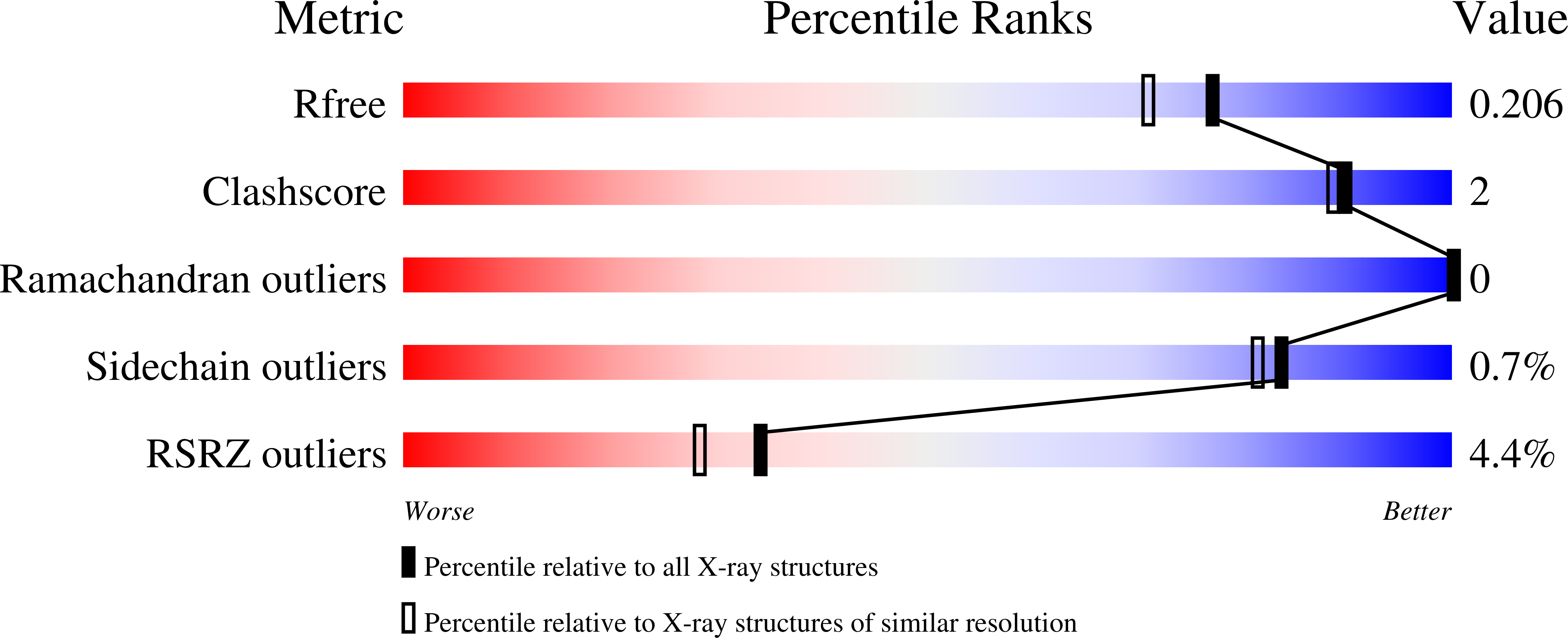
Resolution:
1.79 Å
R-Value Free:
0.20
R-Value Work:
0.17
R-Value Observed:
0.17
Space Group:
P 21 21 21
Macromolecular Entities
Structures with similar UniProt ID
Protein Blast
Polymer Type:polypeptide(L)
Molecule:RE11660p
Gene (Uniprot):phr6-4
Chain IDs:A
Chain Length:502
Number of Molecules:1
Biological Source:Drosophila melanogaster
Primary Citation
The three-dimensional structure of Drosophila melanogaster (6-4) photolyase at room temperature.
Acta Crystallogr D Struct Biol 77 1001 1009 (2021)
PMID: 34342273 DOI: 10.1107/S2059798321005830

Abstact

(6-4) photolyases are flavoproteins that belong to the photolyase/cryptochrome family. Their function is to repair DNA lesions using visible light. Here, crystal structures of Drosophila melanogaster (6-4) photolyase [Dm(6-4)photolyase] at room and cryogenic temperatures are reported. The room-temperature structure was solved to 2.27 Å resolution and was obtained by serial femtosecond crystallography (SFX) using an X-ray free-electron laser. The crystallization and preparation conditions are also reported. The cryogenic structure was solved to 1.79 Å resolution using conventional X-ray crystallography. The structures agree with each other, indicating that the structural information obtained from crystallography at cryogenic temperature also applies at room temperature. Furthermore, UV-Vis absorption spectroscopy confirms that Dm(6-4)photolyase is photoactive in the crystals, giving a green light to time-resolved SFX studies on the protein, which can reveal the structural mechanism of the photoactivated protein in DNA repair.

Legend

Protein

Chemical

Disease

Primary Citation of related structures
Feedback Form
Name
Email
Institute
Feedback