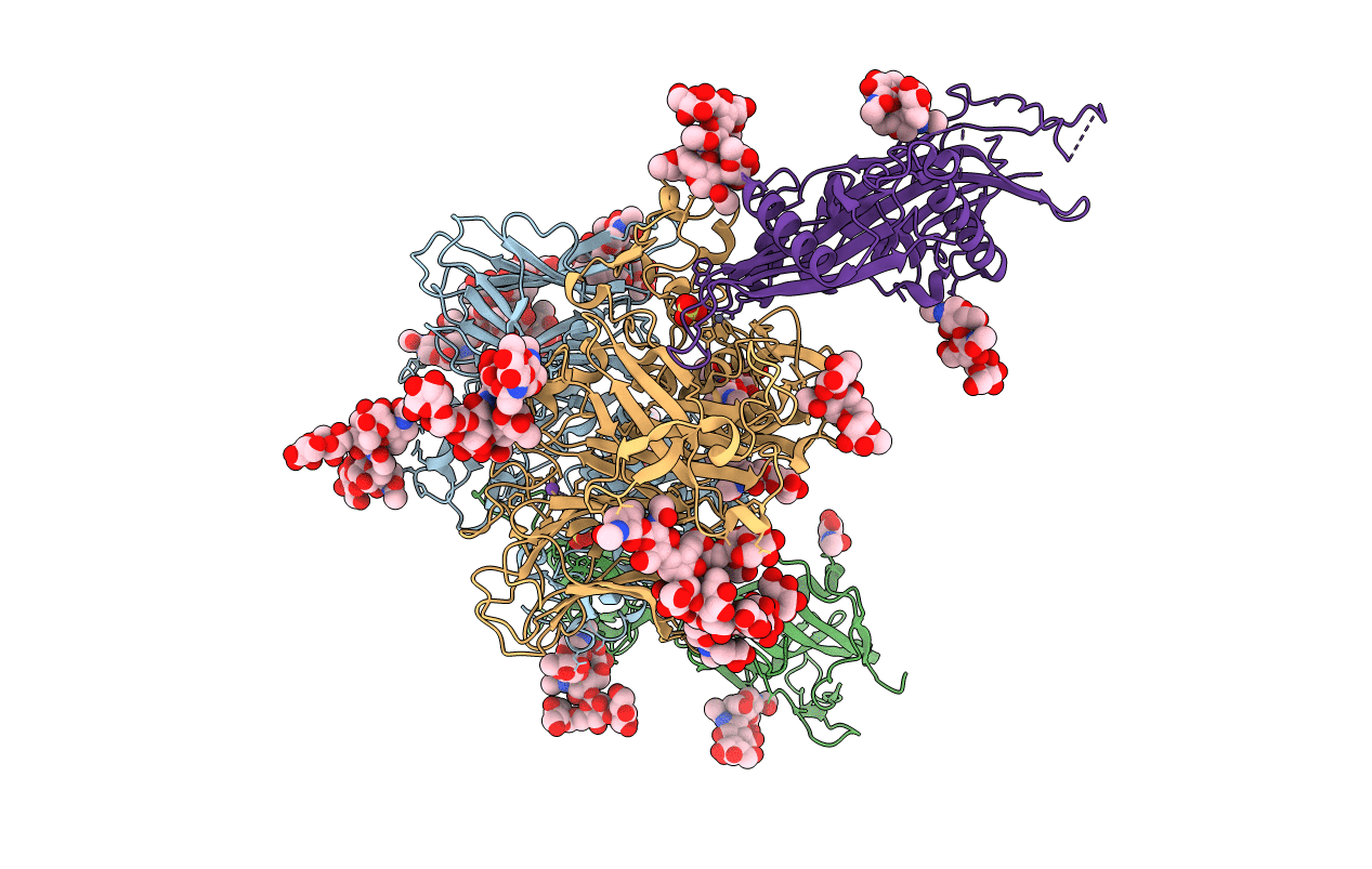
7AUW image
Entry Detail
PDB ID:
7AUW
Title:
Inhibitory complex of human meprin beta with mouse fetuin-B.
Biological Source:
Source Organism:
Host Organism:
PDB Version:
Deposition Date:
2020-11-03
Release Date:
2021-04-14
Method Details:
Experimental Method:
Resolution:
2.80 Å
R-Value Free:
0.26
R-Value Work:
0.23
R-Value Observed:
0.23
Space Group:
P 1 21 1
Macromolecular Entities
Polymer Type:polypeptide(L)
Description:Meprin A subunit beta
Chain IDs:A, C
Chain Length:550
Number of Molecules:2
Biological Source:Homo sapiens
Polymer Type:polypeptide(L)
Description:Fetuin-B
Chain IDs:B, D
Chain Length:394
Number of Molecules:2
Biological Source:Mus musculus
Primary Citation
The crystal structure of a 250-kDa heterotetrameric particle explains inhibition of sheddase meprin beta by endogenous fetuin-B.
Proc.Natl.Acad.Sci.USA 118 ? ? (2021)
PMID: 33782129 DOI: 10.1073/pnas.2023839118

Abstact

Meprin β (Mβ) is a multidomain type-I membrane metallopeptidase that sheds membrane-anchored substrates, releasing their soluble forms. Fetuin-B (FB) is its only known endogenous protein inhibitor. Herein, we analyzed the interaction between the ectodomain of Mβ (MβΔC) and FB, which stabilizes the enzyme and inhibits it with subnanomolar affinity. The MβΔC:FB crystal structure reveals a ∼250-kDa, ∼160-Å polyglycosylated heterotetrameric particle with a remarkable glycan structure. Two FB moieties insert like wedges through a "CPDCP trunk" and two hairpins into the respective peptidase catalytic domains, blocking the catalytic zinc ions through an "aspartate switch" mechanism. Uniquely, the active site clefts are obstructed from subsites S4 to S10', but S1 and S1' are spared, which prevents cleavage. Modeling of full-length Mβ reveals an EGF-like domain between MβΔC and the transmembrane segment that likely serves as a hinge to transit between membrane-distal and membrane-proximal conformations for inhibition and catalysis, respectively.

Legend

Protein

Chemical

Disease

Primary Citation of related structures