Deposition Date
2020-10-21
Release Date
2021-02-24
Last Version Date
2024-11-20
Entry Detail
PDB ID:
7AQG
Keywords:
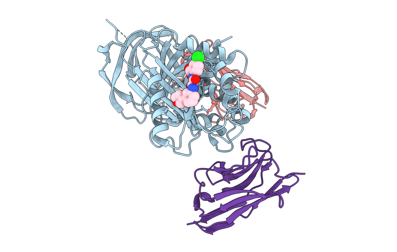
Title:
Crystal Structure of Small Molecule Inhibitor TM5484 Bound to Stabilized Active Plasminogen Activator Inhibitor-1 (PAI-1-W175F)
Biological Source:
Source Organism(s):
Homo sapiens (Taxon ID: 9606)
Vicugna pacos (Taxon ID: 30538)
Vicugna pacos (Taxon ID: 30538)
Expression System(s):
Method Details:
Experimental Method:
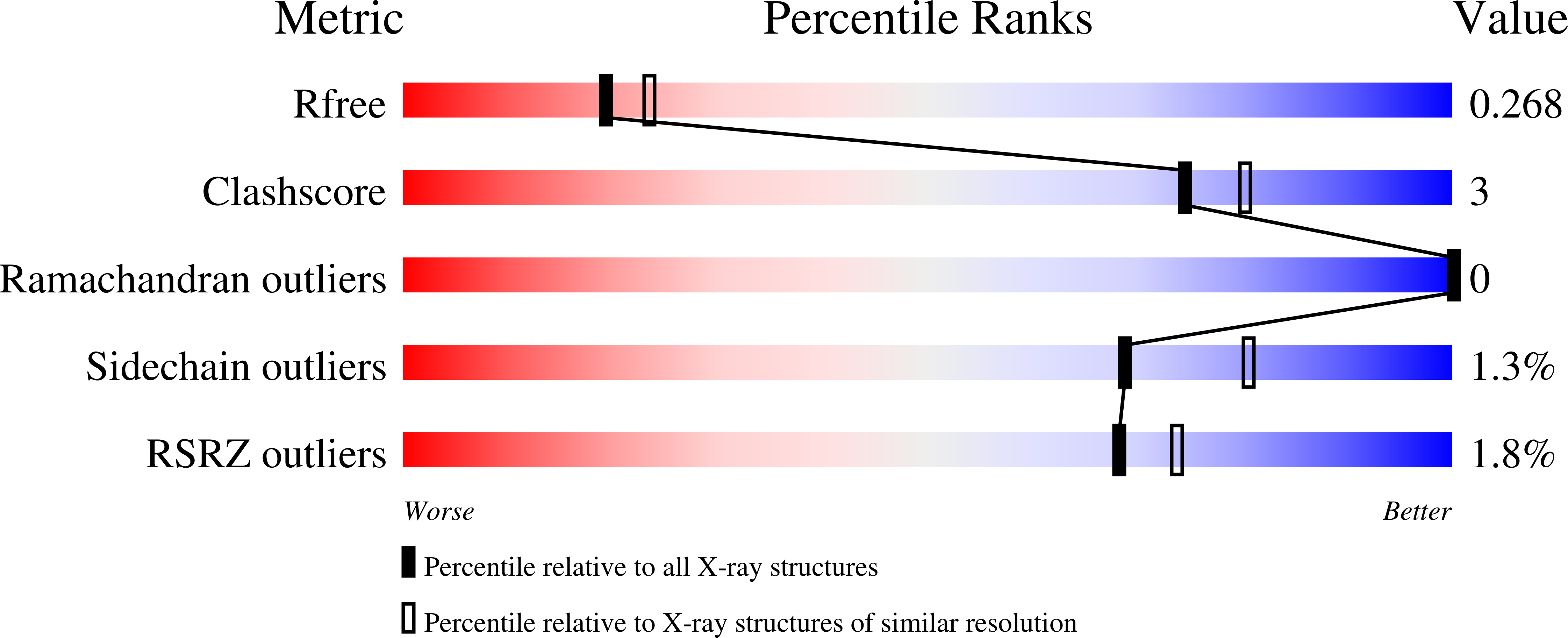
Resolution:
2.27 Å
R-Value Free:
0.26
R-Value Work:
0.21
R-Value Observed:
0.21
Space Group:
P 1 21 1