7AO8 image
Entry Detail
PDB ID:
7AO8
EMDB ID:
Keywords:
Title:
Structure of the MTA1/HDAC1/MBD2 NURD deacetylase complex
Biological Source:
Source Organism:
Host Organism:
PDB Version:
Deposition Date:
2020-10-14
Release Date:
2020-11-11
Method Details:
Experimental Method:
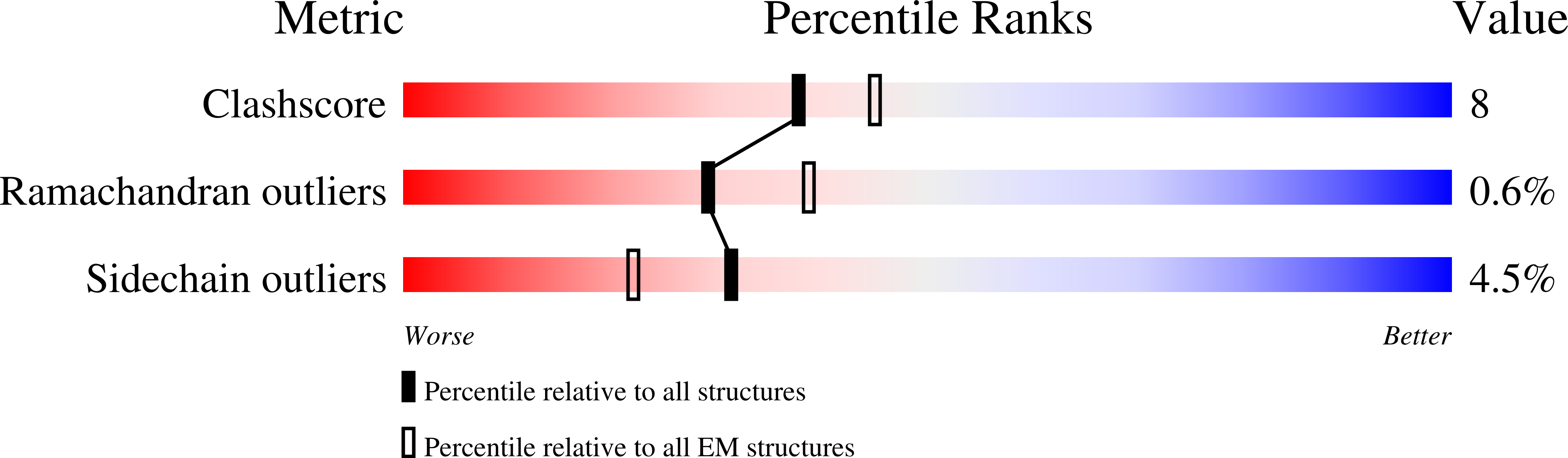
Resolution:
4.50 Å
Aggregation State:
PARTICLE
Reconstruction Method:
SINGLE PARTICLE
Macromolecular Entities
Polymer Type:polypeptide(L)
Description:Methyl-CpG-binding domain protein 2
Chain IDs:A (auth: C)
Chain Length:411
Number of Molecules:1
Biological Source:Homo sapiens
Polymer Type:polypeptide(L)
Description:Metastasis-associated protein MTA1
Chain IDs:B (auth: D), C (auth: A)
Chain Length:715
Number of Molecules:2
Biological Source:Homo sapiens
Polymer Type:polypeptide(L)
Description:Histone deacetylase 1
Chain IDs:D (auth: E), E (auth: B)
Chain Length:482
Number of Molecules:2
Biological Source:Homo sapiens
Primary Citation
The topology of chromatin-binding domains in the NuRD deacetylase complex.
Nucleic Acids Res. 48 12972 12982 (2020)
PMID: 33264408 DOI: 10.1093/nar/gkaa1121

Abstact

Class I histone deacetylase complexes play essential roles in many nuclear processes. Whilst they contain a common catalytic subunit, they have diverse modes of action determined by associated factors in the distinct complexes. The deacetylase module from the NuRD complex contains three protein domains that control the recruitment of chromatin to the deacetylase enzyme, HDAC1/2. Using biochemical approaches and cryo-electron microscopy, we have determined how three chromatin-binding domains (MTA1-BAH, MBD2/3 and RBBP4/7) are assembled in relation to the core complex so as to facilitate interaction of the complex with the genome. We observe a striking arrangement of the BAH domains suggesting a potential mechanism for binding to di-nucleosomes. We also find that the WD40 domains from RBBP4 are linked to the core with surprising flexibility that is likely important for chromatin engagement. A single MBD2 protein binds asymmetrically to the dimerisation interface of the complex. This symmetry mismatch explains the stoichiometry of the complex. Finally, our structures suggest how the holo-NuRD might assemble on a di-nucleosome substrate.

Legend

Protein

Chemical

Disease

Primary Citation of related structures