7AIB image
Entry Detail
PDB ID:
7AIB
EMDB ID:
Title:
MutS-MutL in clamp state
Biological Source:
PDB Version:
Deposition Date:
2020-09-26
Release Date:
2021-03-31
Method Details:
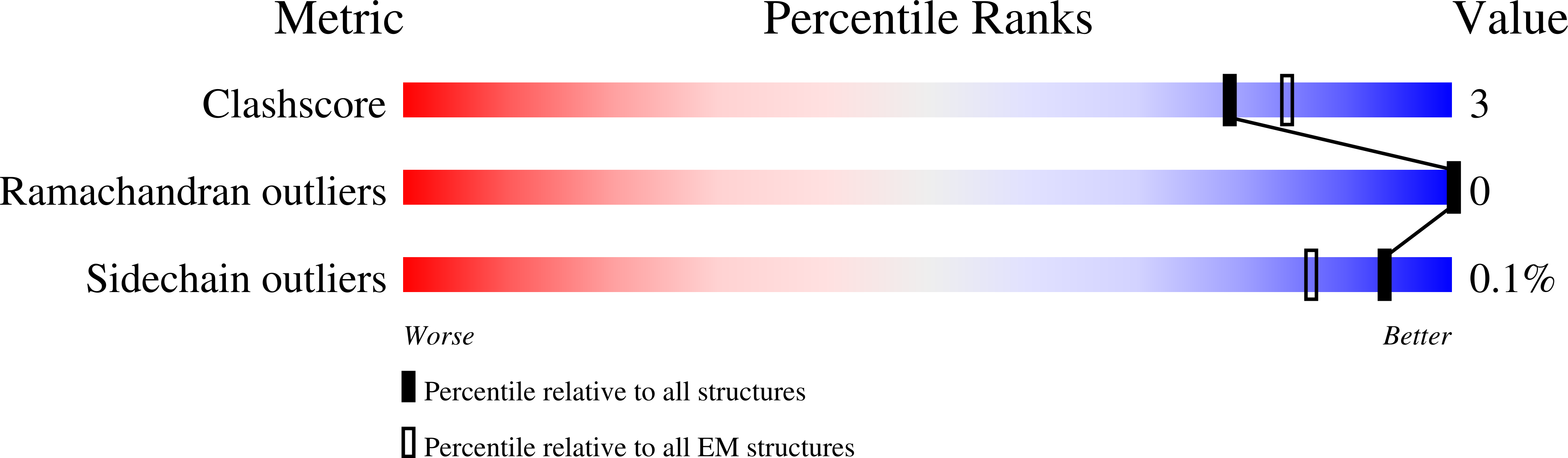
Experimental Method:
Resolution:
4.70 Å
Aggregation State:
PARTICLE
Reconstruction Method:
SINGLE PARTICLE
Macromolecular Entities
Polymer Type:polypeptide(L)
Description:DNA mismatch repair protein MutS
Mutations:D825R,C93A,C235S,C239A,C297S,C569S,C711V,D246C
Chain IDs:A, B
Chain Length:853
Number of Molecules:2
Biological Source:Escherichia coli (strain K12)
Polymer Type:polypeptide(L)
Description:DNA mismatch repair protein MutL
Mutations:N131C,C61S,C216L,C256F,C276Y
Chain IDs:C
Chain Length:351
Number of Molecules:1
Biological Source:Escherichia coli (strain K12)
Polymer Type:polydeoxyribonucleotide
Description:DNA (30-MER)
Chain IDs:D
Chain Length:30
Number of Molecules:1
Biological Source:synthetic construct
Polymer Type:polydeoxyribonucleotide
Description:DNA (30-MER)
Chain IDs:E
Chain Length:30
Number of Molecules:1
Biological Source:synthetic construct
Ligand Molecules
Primary Citation
The selection process of licensing a DNA mismatch for repair.
Nat.Struct.Mol.Biol. 28 373 381 (2021)
PMID: 33820992 DOI: 10.1038/s41594-021-00577-7

Abstact

DNA mismatch repair detects and removes mismatches from DNA by a conserved mechanism, reducing the error rate of DNA replication by 100- to 1,000-fold. In this process, MutS homologs scan DNA, recognize mismatches and initiate repair. How the MutS homologs selectively license repair of a mismatch among millions of matched base pairs is not understood. Here we present four cryo-EM structures of Escherichia coli MutS that provide snapshots, from scanning homoduplex DNA to mismatch binding and MutL activation via an intermediate state. During scanning, the homoduplex DNA forms a steric block that prevents MutS from transitioning into the MutL-bound clamp state, which can only be overcome through kinking of the DNA at a mismatch. Structural asymmetry in all four structures indicates a division of labor between the two MutS monomers. Together, these structures reveal how a small conformational change from the homoduplex- to heteroduplex-bound MutS acts as a licensing step that triggers a dramatic conformational change that enables MutL binding and initiation of the repair cascade.

Legend

Protein

Chemical

Disease

Primary Citation of related structures