7AED image
Deposition Date 2020-09-17
Release Date 2020-11-18
Last Version Date 2024-06-19
Entry Detail
PDB ID:
7AED
Title:
VirB8 domain of PrgL from Enterococcus faecalis pCF10
Biological Source:
Source Organism:
Host Organism:
Method Details:
Experimental Method:
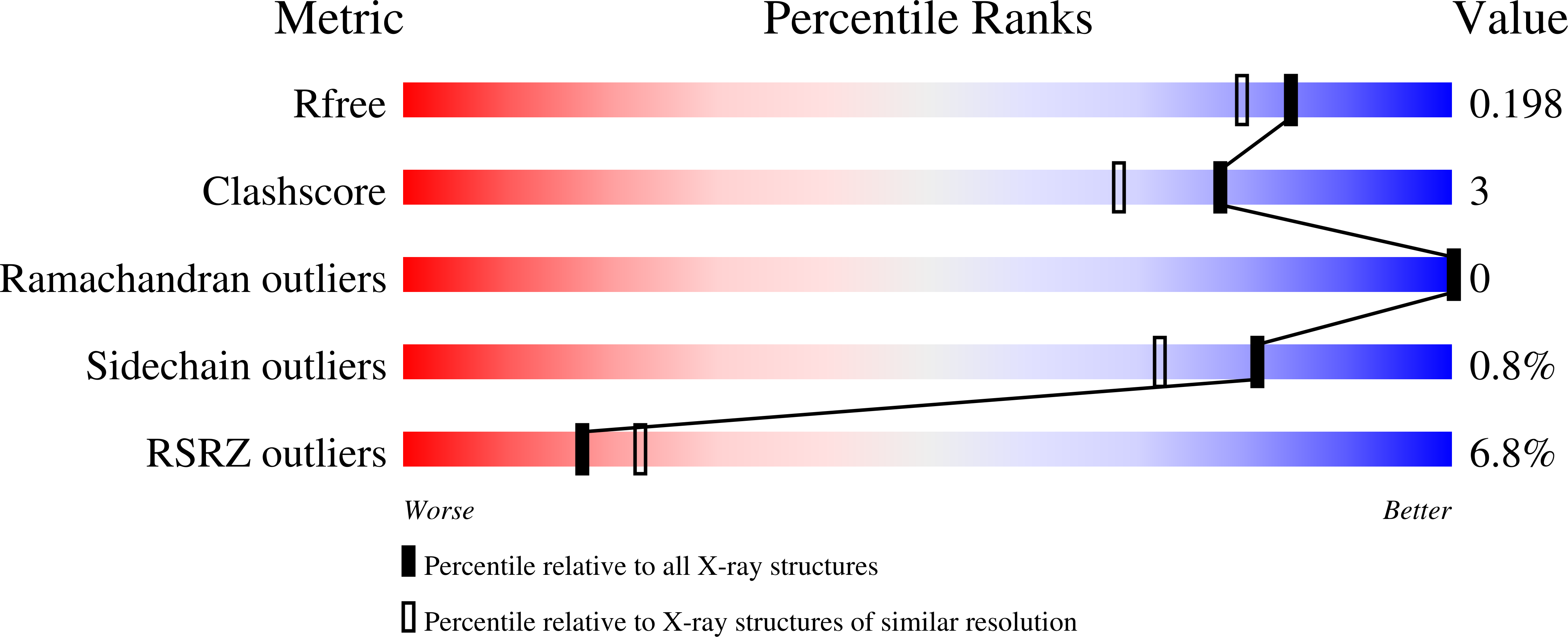
Resolution:
1.75 Å
R-Value Free:
0.19
R-Value Work:
0.17
R-Value Observed:
0.18
Space Group:
H 3
Macromolecular Entities
Structures with similar UniProt ID
Protein Blast
Polymer Type:polypeptide(L)
Molecule:PrgL
Gene (Uniprot):prgL
Chain IDs:A, B
Chain Length:195
Number of Molecules:2
Biological Source:Enterococcus faecalis
Ligand Molecules
Primary Citation
Structure of the enterococcal T4SS protein PrgL reveals unique dimerization interface in the VirB8 protein family.
Structure 30 876 ? (2022)
PMID: 35429437 DOI: 10.1016/j.str.2022.03.013

Abstact

Multidrug-resistant bacteria pose serious problems in hospital-acquired infections (HAIs). Most antibiotic resistance genes are acquired via conjugative gene transfer, mediated by type 4 secretion systems (T4SS). Although most multidrug-resistant bacteria responsible for HAIs are of Gram-positive origin, with enterococci being major contributors, mostly Gram-negative T4SSs have been characterized. Here, we describe the structure and organization of PrgL, a core protein of the T4SS channel, encoded by the pCF10 plasmid from Enterococcus faecalis. The structure of PrgL displays similarity to VirB8 proteins of Gram-negative T4SSs. In vitro experiments show that the soluble domain alone is enough to drive both dimerization and dodecamerization, with a dimerization interface that differs from all other known VirB8-like proteins. In vivo experiments verify the importance of PrgL dimerization. Our findings provide insight into the molecular building blocks of Gram-positive T4SS, highlighting similarities but also unique features in PrgL compared to other VirB8-like proteins.

Legend

Protein

Chemical

Disease

Primary Citation of related structures