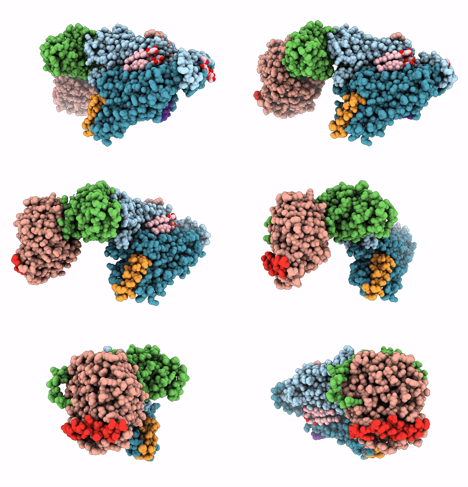
7AD3 image
Entry Detail
PDB ID:
7AD3
EMDB ID:
Title:
Class D GPCR Ste2 dimer coupled to two G proteins
Biological Source:
PDB Version:
Deposition Date:
2020-09-14
Release Date:
2020-12-09
Method Details:
Experimental Method:
Resolution:
3.00 Å
Aggregation State:
PARTICLE
Reconstruction Method:
SINGLE PARTICLE
Macromolecular Entities
Polymer Type:polypeptide(L)
Description:Pheromone alpha factor receptor
Chain IDs:A (auth: B), B (auth: A)
Chain Length:431
Number of Molecules:2
Biological Source:Saccharomyces cerevisiae
Polymer Type:polypeptide(L)
Description:Guanine nucleotide-binding protein alpha-1 subunit,Guanine nucleotide-binding protein alpha-1 subunit
Chain IDs:F (auth: E), H
Chain Length:233
Number of Molecules:2
Biological Source:Saccharomyces cerevisiae (strain ATCC 204508 / S288c)
Polymer Type:polypeptide(L)
Description:STE4 isoform 1
Chain IDs:E (auth: F)
Chain Length:423
Number of Molecules:1
Biological Source:Saccharomyces cerevisiae
Polymer Type:polypeptide(L)
Description:Guanine nucleotide-binding protein subunit gamma
Chain IDs:G
Chain Length:109
Number of Molecules:1
Biological Source:Saccharomyces cerevisiae
Polymer Type:polypeptide(L)
Description:Alpha-factor mating pheromone
Chain IDs:C (auth: K), D (auth: I)
Chain Length:13
Number of Molecules:2
Biological Source:Saccharomyces cerevisiae
Primary Citation
Structure of the class D GPCR Ste2 dimer coupled to two G proteins.
Nature 589 148 153 (2021)
PMID: 33268889 DOI: 10.1038/s41586-020-2994-1

Abstact

G-protein-coupled receptors (GPCRs) are divided phylogenetically into six classes1,2, denoted A to F. More than 370 structures of vertebrate GPCRs (belonging to classes A, B, C and F) have been determined, leading to a substantial understanding of their function3. By contrast, there are no structures of class D GPCRs, which are found exclusively in fungi where they regulate survival and reproduction. Here we determine the structure of a class D GPCR, the Saccharomyces cerevisiae pheromone receptor Ste2, in an active state coupled to the heterotrimeric G protein Gpa1-Ste4-Ste18. Ste2 was purified as a homodimer coupled to two G proteins. The dimer interface of Ste2 is formed by the N terminus, the transmembrane helices H1, H2 and H7, and the first extracellular loop ECL1. We establish a class D1 generic residue numbering system (CD1) to enable comparisons with orthologues and with other GPCR classes. The structure of Ste2 bears similarities in overall topology to class A GPCRs, but the transmembrane helix H4 is shifted by more than 20 Å and the G-protein-binding site is a shallow groove rather than a cleft. The structure provides a template for the design of novel drugs to target fungal GPCRs, which could be used to treat numerous intractable fungal diseases4.

Legend

Protein

Chemical

Disease

Primary Citation of related structures