7ABW image
Entry Detail
PDB ID:
7ABW
Title:
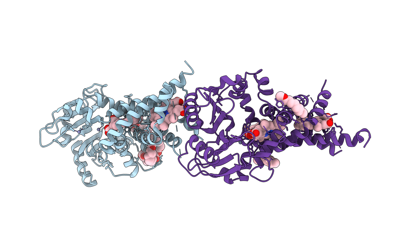
Crystal structure of siderophore reductase FoxB
Biological Source:
Source Organism:
Host Organism:
PDB Version:
Deposition Date:
2020-09-09
Release Date:
2021-09-01
Method Details:
Experimental Method:
Resolution:
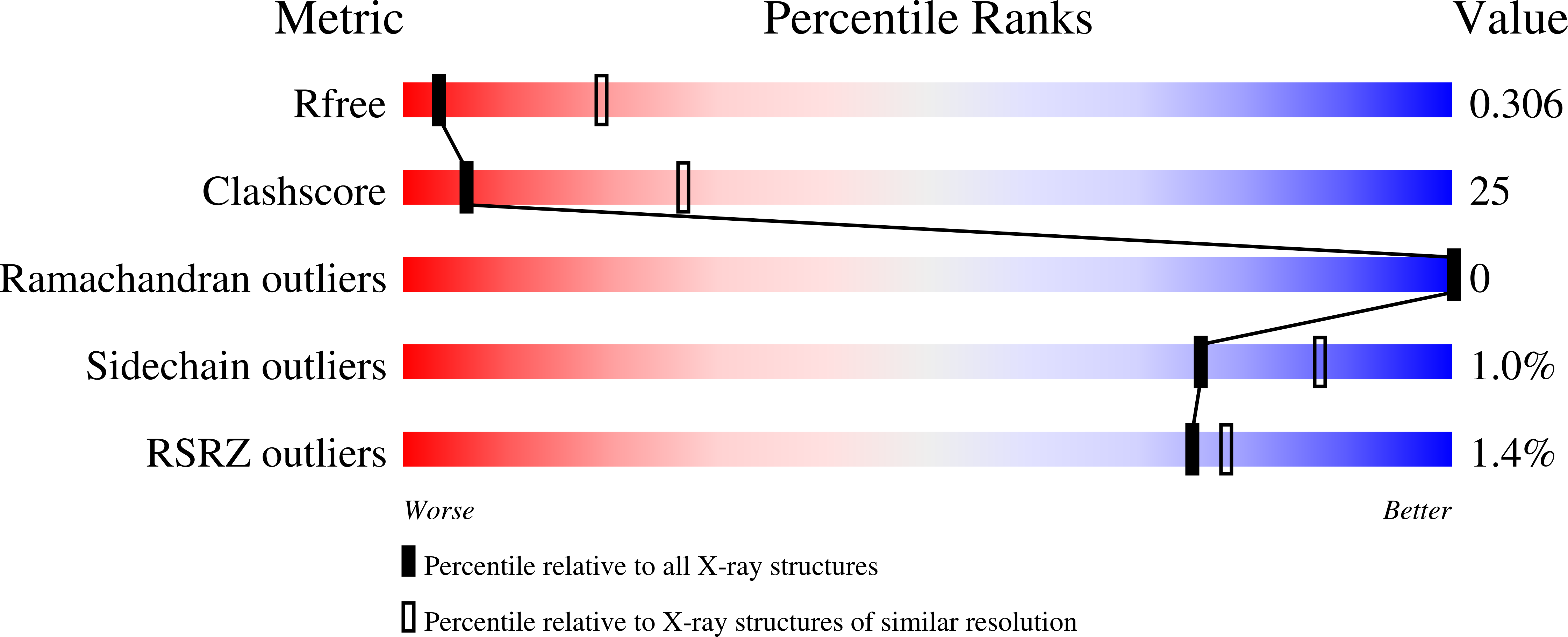
3.35 Å
R-Value Free:
0.29
R-Value Work:
0.26
R-Value Observed:
0.26
Space Group:
P 21 21 2
Macromolecular Entities
Polymer Type:polypeptide(L)
Description:PepSY domain-containing protein
Chain IDs:A, B
Chain Length:382
Number of Molecules:2
Biological Source:Pseudomonas aeruginosa
Primary Citation
Structural insights into a novel family of integral membrane siderophore reductases.
Proc.Natl.Acad.Sci.USA 118 ? ? (2021)
PMID: 34417315 DOI: 10.1073/pnas.2101952118

Abstact

Gram-negative bacteria take up the essential ion Fe3+ as ferric-siderophore complexes through their outer membrane using TonB-dependent transporters. However, the subsequent route through the inner membrane differs across many bacterial species and siderophore chemistries and is not understood in detail. Here, we report the crystal structure of the inner membrane protein FoxB (from Pseudomonas aeruginosa) that is involved in Fe-siderophore uptake. The structure revealed a fold with two tightly bound heme molecules. In combination with in vitro reduction assays and in vivo iron uptake studies, these results establish FoxB as an inner membrane reductase involved in the release of iron from ferrioxamine during Fe-siderophore uptake.

Legend

Protein

Chemical

Disease

Primary Citation of related structures