7ABM image
Entry Detail
PDB ID:
7ABM
Title:
X-ray structure of phosphorylated Barrier-to-autointegration factor (BAF)
Biological Source:
Source Organism:
Host Organism:
PDB Version:
Deposition Date:
2020-09-08
Release Date:
2021-07-07
Method Details:
Experimental Method:
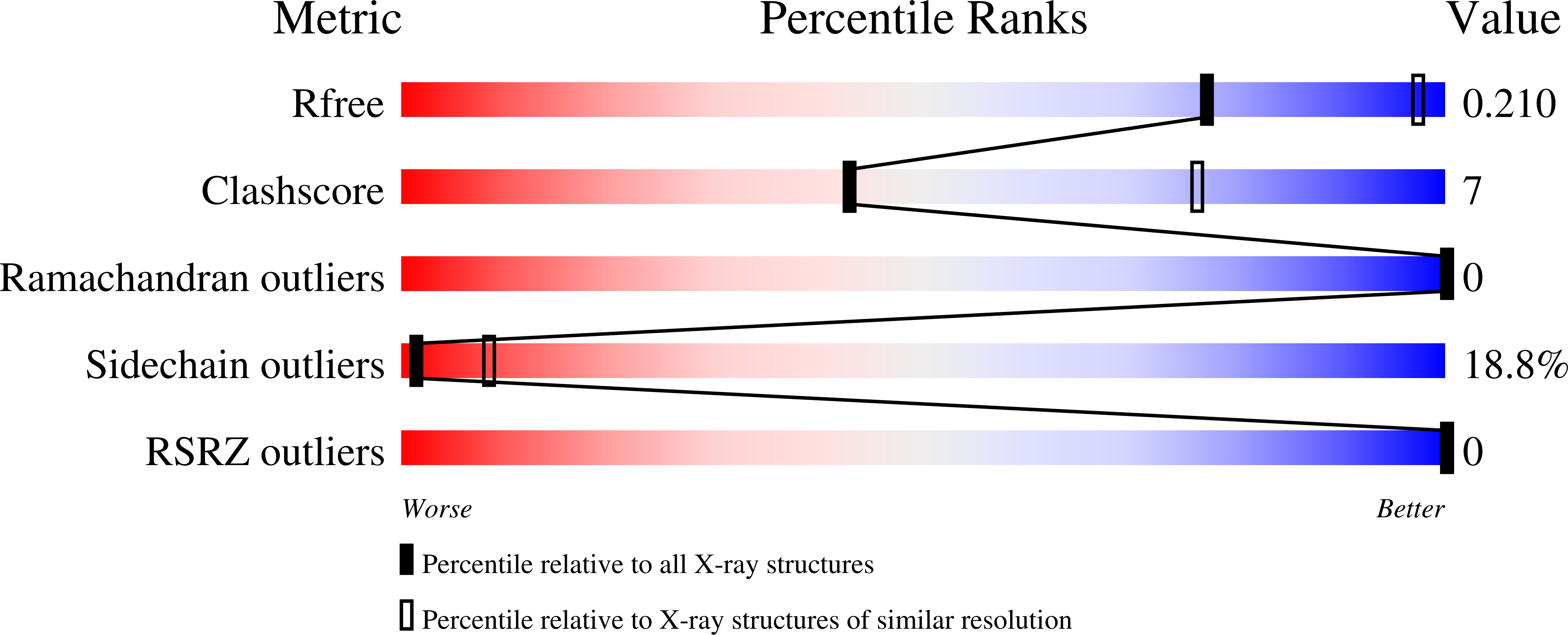
Resolution:
3.00 Å
R-Value Free:
0.32
R-Value Work:
0.19
R-Value Observed:
0.20
Space Group:
P 32 2 1
Macromolecular Entities
Polymer Type:polypeptide(L)
Description:Barrier-to-autointegration factor
Chain IDs:A
Chain Length:86
Number of Molecules:1
Biological Source:Homo sapiens
Modified Residue
Compound ID Chain ID Parent Comp ID Details 2D Image
SEP A SER modified residue
Ligand Molecules
Primary Citation
Di-phosphorylated BAF shows altered structural dynamics and binding to DNA, but interacts with its nuclear envelope partners.
Nucleic Acids Res. 49 3841 3855 (2021)
PMID: 33744941 DOI: 10.1093/nar/gkab184

Abstact

Barrier-to-autointegration factor (BAF), encoded by the BANF1 gene, is an abundant and ubiquitously expressed metazoan protein that has multiple functions during the cell cycle. Through its ability to cross-bridge two double-stranded DNA (dsDNA), it favours chromosome compaction, participates in post-mitotic nuclear envelope reassembly and is essential for the repair of large nuclear ruptures. BAF forms a ternary complex with the nuclear envelope proteins lamin A/C and emerin, and its interaction with lamin A/C is defective in patients with recessive accelerated aging syndromes. Phosphorylation of BAF by the vaccinia-related kinase 1 (VRK1) is a key regulator of BAF localization and function. Here, we demonstrate that VRK1 successively phosphorylates BAF on Ser4 and Thr3. The crystal structures of BAF before and after phosphorylation are extremely similar. However, in solution, the extensive flexibility of the N-terminal helix α1 and loop α1α2 in BAF is strongly reduced in di-phosphorylated BAF, due to interactions between the phosphorylated residues and the positively charged C-terminal helix α6. These regions are involved in DNA and lamin A/C binding. Consistently, phosphorylation causes a 5000-fold loss of affinity for dsDNA. However, it does not impair binding to lamin A/C Igfold domain and emerin nucleoplasmic region, which leaves open the question of the regulation of these interactions.

Legend

Protein

Chemical

Disease

Primary Citation of related structures