Deposition Date
2020-08-31
Release Date
2021-10-06
Last Version Date
2024-11-06
Entry Detail
PDB ID:
7A8Y
Keywords:
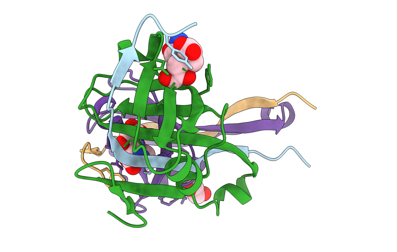
Title:
X-ray crystal structure of Aspartate alpha-decarboxylase in complex with D-Serine
Biological Source:
Source Organism(s):
Escherichia coli (Taxon ID: 562)
Expression System(s):
Method Details:
Experimental Method:
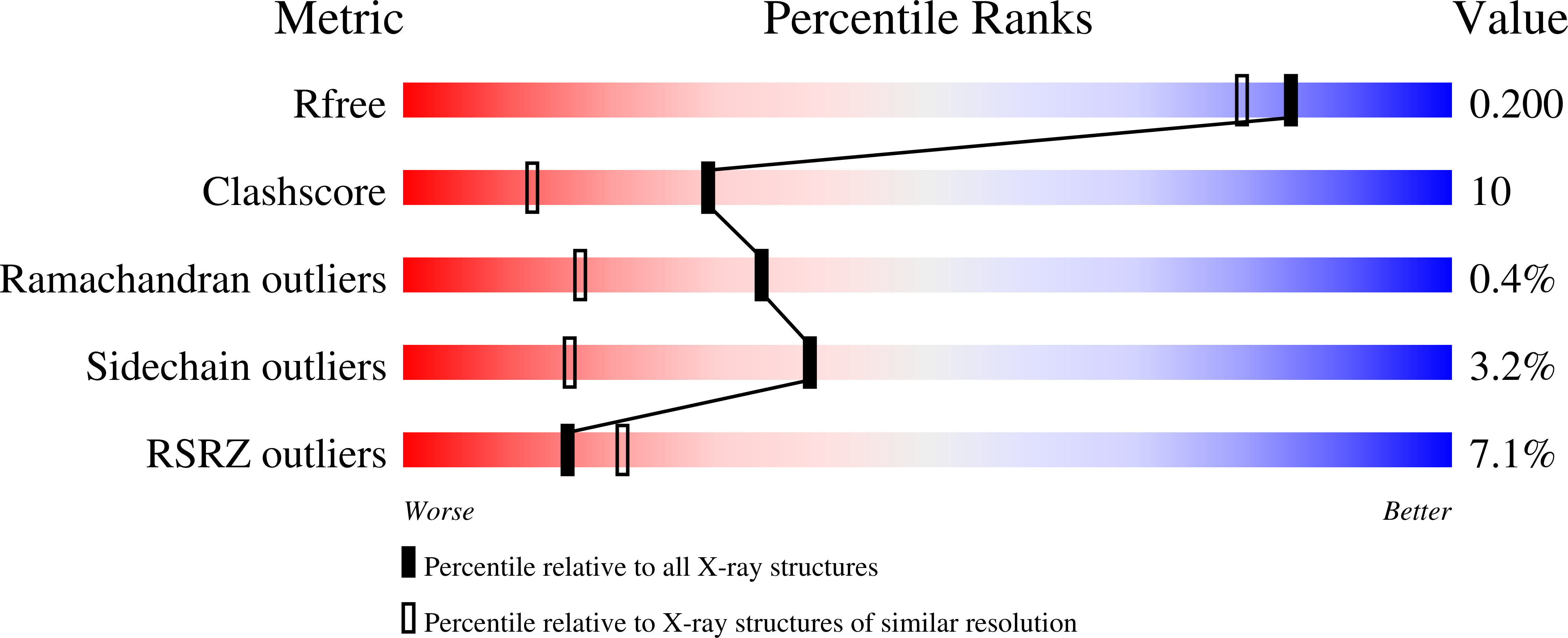
Resolution:
1.75 Å
R-Value Free:
0.19
R-Value Work:
0.17
Space Group:
P 61 2 2