
Deposition Date
2019-04-19
Release Date
2019-05-15
Last Version Date
2024-05-15
Entry Detail
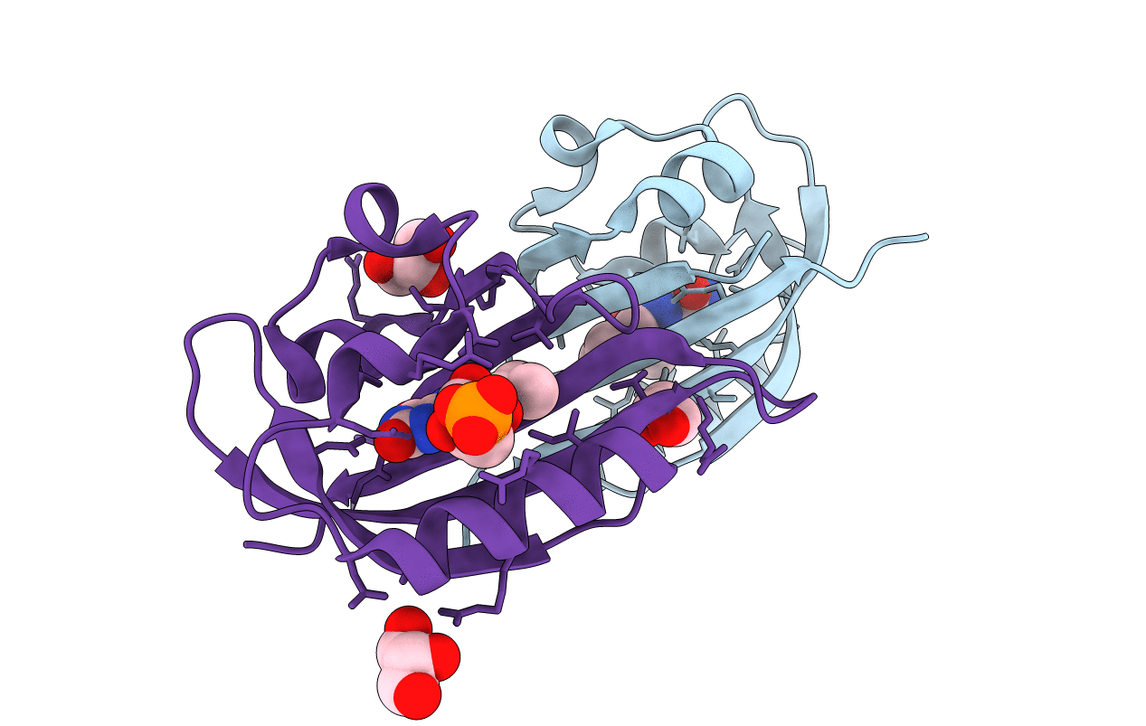
PDB ID:
6RHF
Keywords:
Title:
Structure of Chloroflexus aggregans Cagg_3753 LOV domain C85A variant (CagFbFP)
Biological Source:
Source Organism(s):
Expression System(s):
Method Details:
Experimental Method:
Resolution:
1.07 Å
R-Value Free:
0.14
R-Value Work:
0.12
R-Value Observed:
0.12
Space Group:
P 21 21 2
