
Deposition Date
2017-12-10
Release Date
2018-10-10
Last Version Date
2024-11-20
Entry Detail
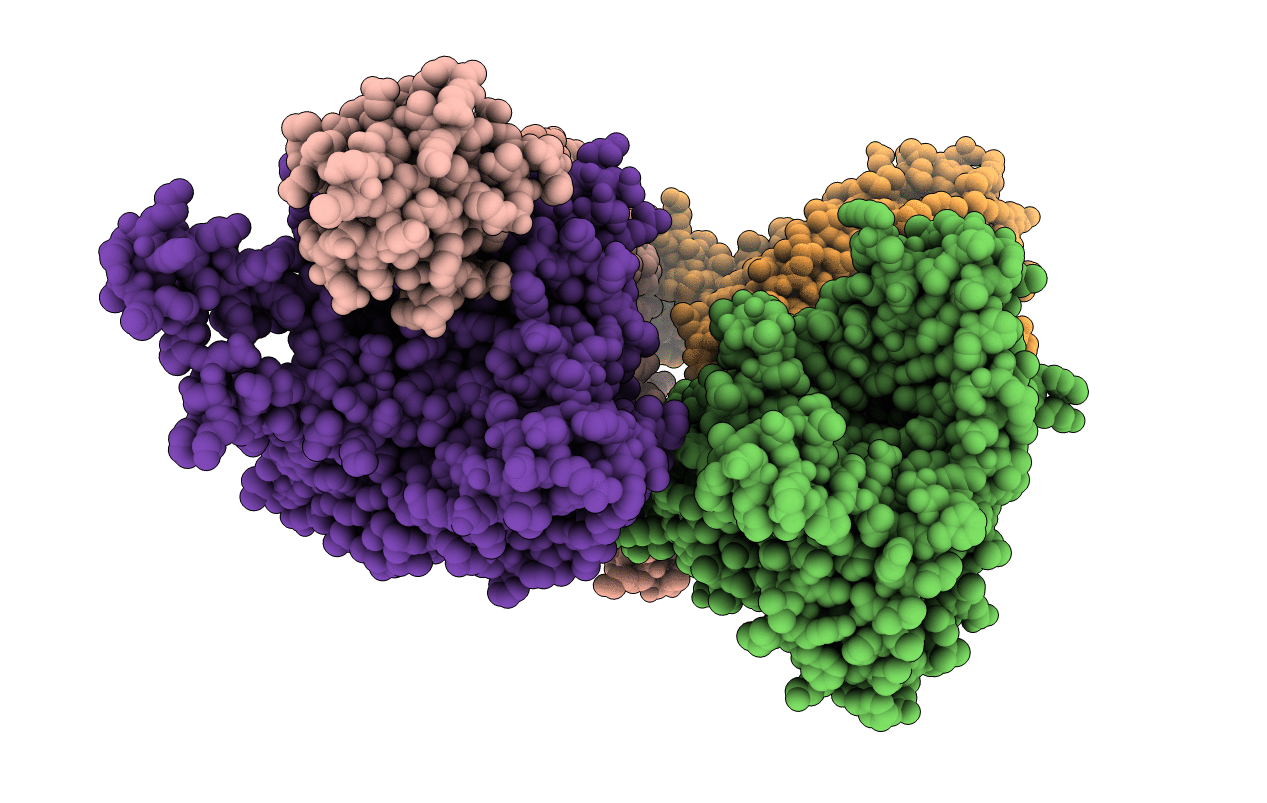
PDB ID:
6BUH
Keywords:
Title:
Crystal structure of a membrane protein, crystal form II
Biological Source:
Source Organism(s):
Streptococcus thermophilus (Taxon ID: 1308)
Expression System(s):
Method Details:
Experimental Method:
Resolution:
3.15 Å
R-Value Free:
0.29
R-Value Work:
0.27
R-Value Observed:
0.27
Space Group:
P 1 21 1
