6ZWK image
Entry Detail
PDB ID:
6ZWK
Keywords:
Title:
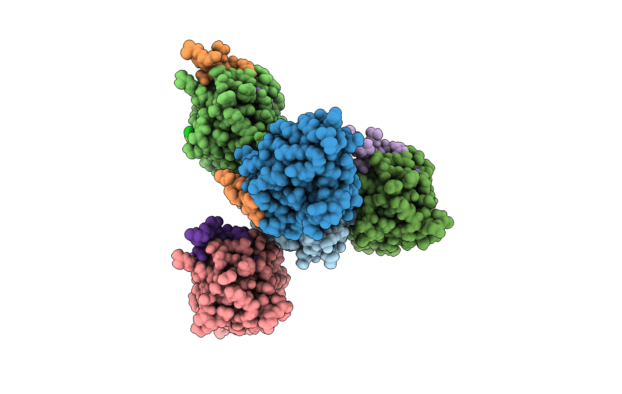
Crystal structure of the phosphorylated C-terminal tail of histone H2AX in complex with a specific nanobody (C6 gammaXbody)
Biological Source:
Source Organism:
PDB Version:
Deposition Date:
2020-07-28
Release Date:
2021-07-21
Method Details:
Experimental Method:
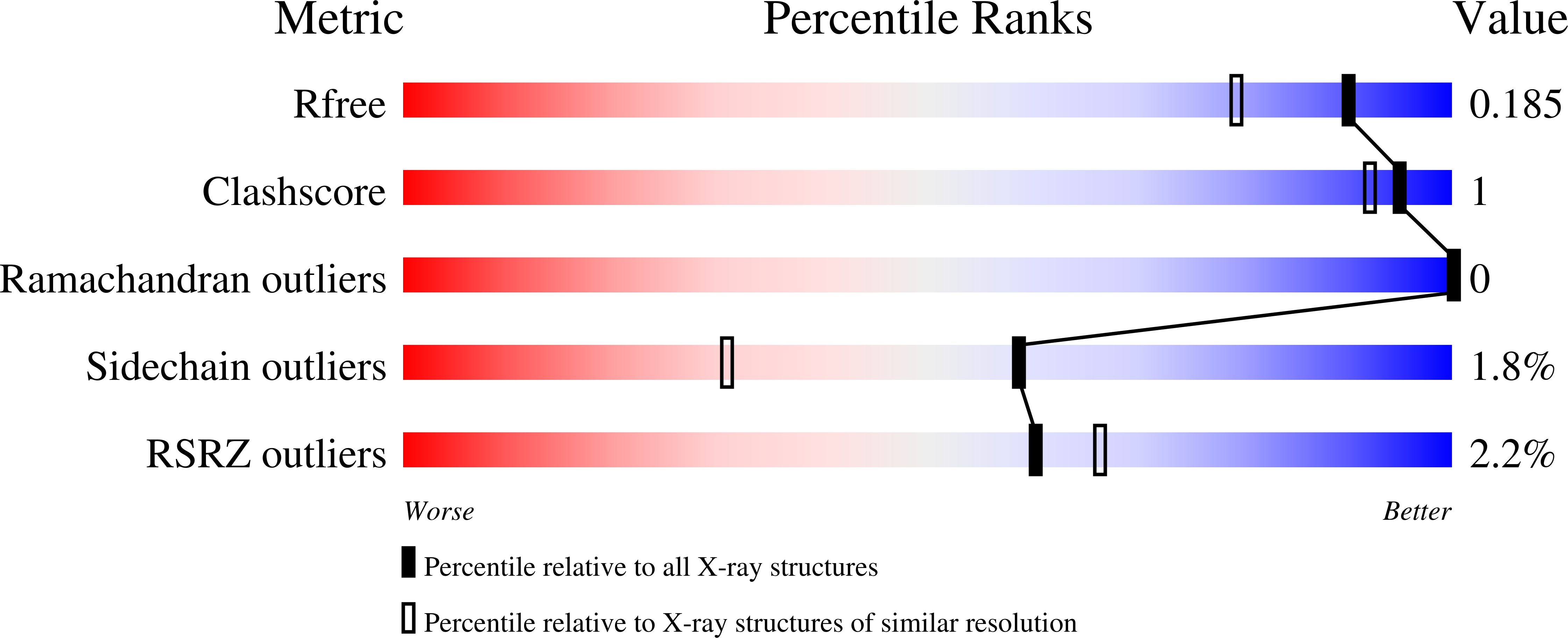
Resolution:
1.55 Å
R-Value Free:
0.18
R-Value Work:
0.14
R-Value Observed:
0.15
Space Group:
P 31
Macromolecular Entities
Polymer Type:polypeptide(L)
Description:gammaXbody
Chain IDs:A, B, C, D, E, F
Chain Length:150
Number of Molecules:6
Biological Source:Vicugna pacos
Polymer Type:polypeptide(L)
Description:Histone H2AX
Chain IDs:G, H, I, J, K, L
Chain Length:10
Number of Molecules:6
Biological Source:Homo sapiens
Modified Residue
Compound ID Chain ID Parent Comp ID Details 2D Image
SEP G SER modified residue
Primary Citation
A Novel Nanobody Precisely Visualizes Phosphorylated Histone H2AX in Living Cancer Cells under Drug-Induced Replication Stress.
Cancers (Basel) 13 ? ? (2021)
PMID: 34282773 DOI: 10.3390/cancers13133317

Abstact

Histone H2AX phosphorylated at serine 139 (γ-H2AX) is a hallmark of DNA damage, signaling the presence of DNA double-strand breaks and global replication stress in mammalian cells. While γ-H2AX can be visualized with antibodies in fixed cells, its detection in living cells was so far not possible. Here, we used immune libraries and phage display to isolate nanobodies that specifically bind to γ-H2AX. We solved the crystal structure of the most soluble nanobody in complex with the phosphopeptide corresponding to the C-terminus of γ-H2AX and show the atomic constituents behind its specificity. We engineered a bivalent version of this nanobody and show that bivalency is essential to quantitatively visualize γ-H2AX in fixed drug-treated cells. After labelling with a chemical fluorophore, we were able to detect γ-H2AX in a single-step assay with the same sensitivity as with validated antibodies. Moreover, we produced fluorescent nanobody-dTomato fusion proteins and applied a transduction strategy to visualize with precision γ-H2AX foci present in intact living cells following drug treatment. Together, this novel tool allows performing fast screenings of genotoxic drugs and enables to study the dynamics of this particular chromatin modification in individual cancer cells under a variety of conditions.

Legend

Protein

Chemical

Disease

Primary Citation of related structures