Deposition Date
2020-07-25
Release Date
2020-10-14
Last Version Date
2024-01-31
Entry Detail
PDB ID:
6ZVN
Keywords:
Title:
Botulinum neurotoxin B2 binding domain in complex with human synaptotagmin I
Biological Source:
Source Organism:
Clostridium botulinum (Taxon ID: 1491)
Homo sapiens (Taxon ID: 9606)
Homo sapiens (Taxon ID: 9606)
Host Organism:
Method Details:
Experimental Method:
Resolution:
2.50 Å
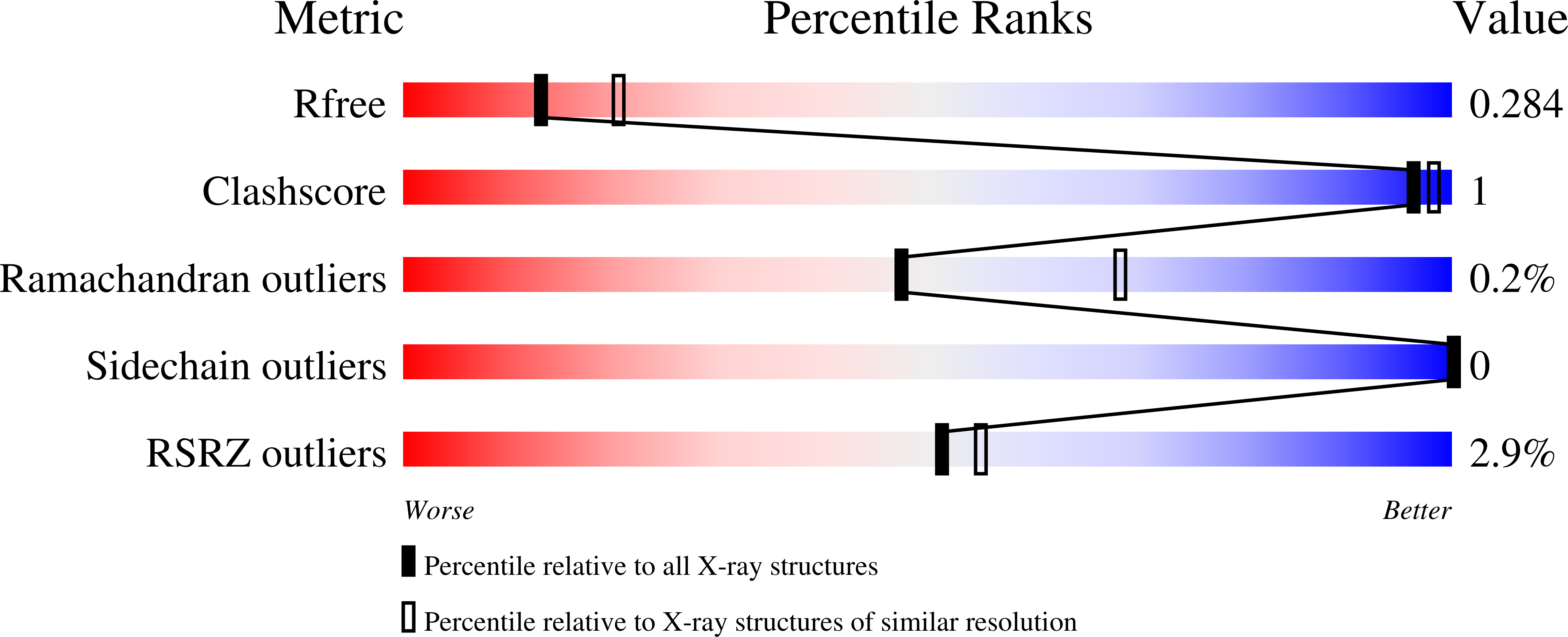
R-Value Free:
0.28
R-Value Work:
0.25
Space Group:
P 21 21 21