6ZVF image
Deposition Date 2020-07-24
Release Date 2021-04-14
Last Version Date 2024-11-20
Entry Detail
PDB ID:
6ZVF
Keywords:
Title:
Crystal structure of the recombinant Fab fragment derived from the hybridoma M3/38 in complex with a human Galectin-3 peptide
Biological Source:
Source Organism:
Rattus norvegicus (Taxon ID: 10116)
Homo sapiens (Taxon ID: 9606)
Host Organism:
Method Details:
Experimental Method:
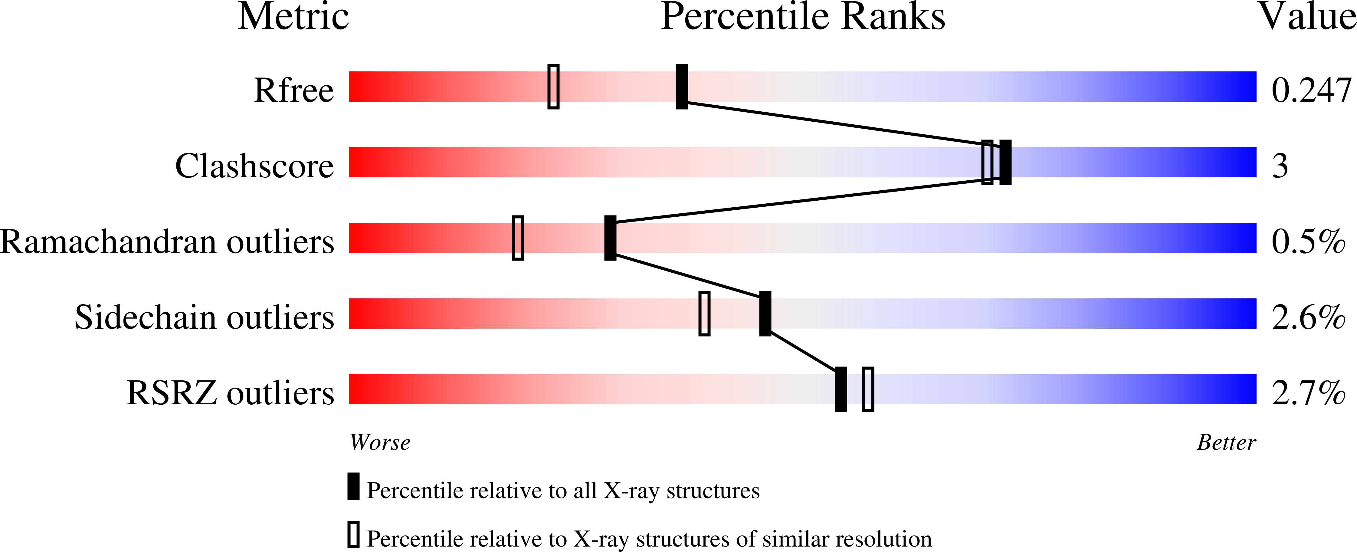
Resolution:
1.90 Å
R-Value Free:
0.24
R-Value Work:
0.20
R-Value Observed:
0.20
Space Group:
C 1 2 1
Macromolecular Entities
Polymer Type:polypeptide(L)
Molecule:Chimeric Fab M3/38 (L,H)
Chain IDs:B (auth: H)
Chain Length:227
Number of Molecules:1
Biological Source:Rattus norvegicus
Polymer Type:polypeptide(L)
Molecule:Chimeric Fab M3/38 (L,H)
Chain IDs:A (auth: L)
Chain Length:219
Number of Molecules:1
Biological Source:Rattus norvegicus
Structures with similar UniProt ID
Protein Blast
Polymer Type:polypeptide(L)
Molecule:Galectin-3
Gene (Uniprot):LGALS3
Chain IDs:C (auth: P)
Chain Length:10
Number of Molecules:1
Biological Source:Homo sapiens
Primary Citation
Effective rational humanization of a PASylated anti-galectin-3 Fab for the sensitive PET imaging of thyroid cancer in vivo.
Sci Rep 11 7358 7358 (2021)
PMID: 33795750 DOI: 10.1038/s41598-021-86641-0

Abstact

The lack of a non-invasive test for malignant thyroid nodules makes the diagnosis of thyroid cancer (TC) challenging. Human galectin-3 (hGal3) has emerged as a promising target for medical TC imaging and diagnosis because of its exclusive overexpression in malignant thyroid tissues. We previously developed a human-chimeric αhGal3 Fab fragment derived from the rat monoclonal antibody (mAb) M3/38 with optimized clearance characteristics using PASylation technology. Here, we describe the elucidation of the hGal3 epitope recognized by mAb M3/38, X-ray crystallographic analysis of its complex with the chimeric Fab and, based on the three-dimensional structure, the rational humanization of the Fab by CDR grafting. Four CDR-grafted versions were designed using structurally most closely related fully human immunoglobulin VH/VL regions of which one-employing the acceptor framework regions of the HIV-1 neutralizing human antibody m66-showed the highest antigen affinity. By introducing two additional back-mutations to the rodent donor sequence, an affinity toward hGal3 indistinguishable from the chimeric Fab was achieved (KD = 0.34 ± 0.02 nM in SPR). The PASylated humanized Fab was site-specifically labelled with the fluorescent dye Cy7 and applied for the immuno-histochemical staining of human tissue sections representative for different TCs. The same protein was conjugated with the metal chelator Dfo, followed by radiolabelling with 89Zr(IV). The resulting protein tracer allowed the highly sensitive and specific PET/CT imaging of orthotopic tumors in mice, which was confirmed by quantitative analysis of radiotracer accumulation. Thus, the PASylated humanized αhGal3 Fab offers clinical potential for the diagnostic imaging of TC.

Legend

Protein

Chemical

Disease

Primary Citation of related structures
Feedback Form
Name
Email
Institute
Feedback