Deposition Date
2020-07-23
Release Date
2021-06-30
Last Version Date
2024-10-16
Entry Detail
PDB ID:
6ZUI
Keywords:
Title:
Crystal structure of the Cys-Ser mutant of the cpYFP-based biosensor for hypochlorous acid
Biological Source:
Source Organism(s):
Escherichia coli (strain K12) (Taxon ID: 83333)
Aequorea victoria (Taxon ID: 6100)
Aequorea victoria (Taxon ID: 6100)
Expression System(s):
Method Details:
Experimental Method:
Resolution:
2.20 Å
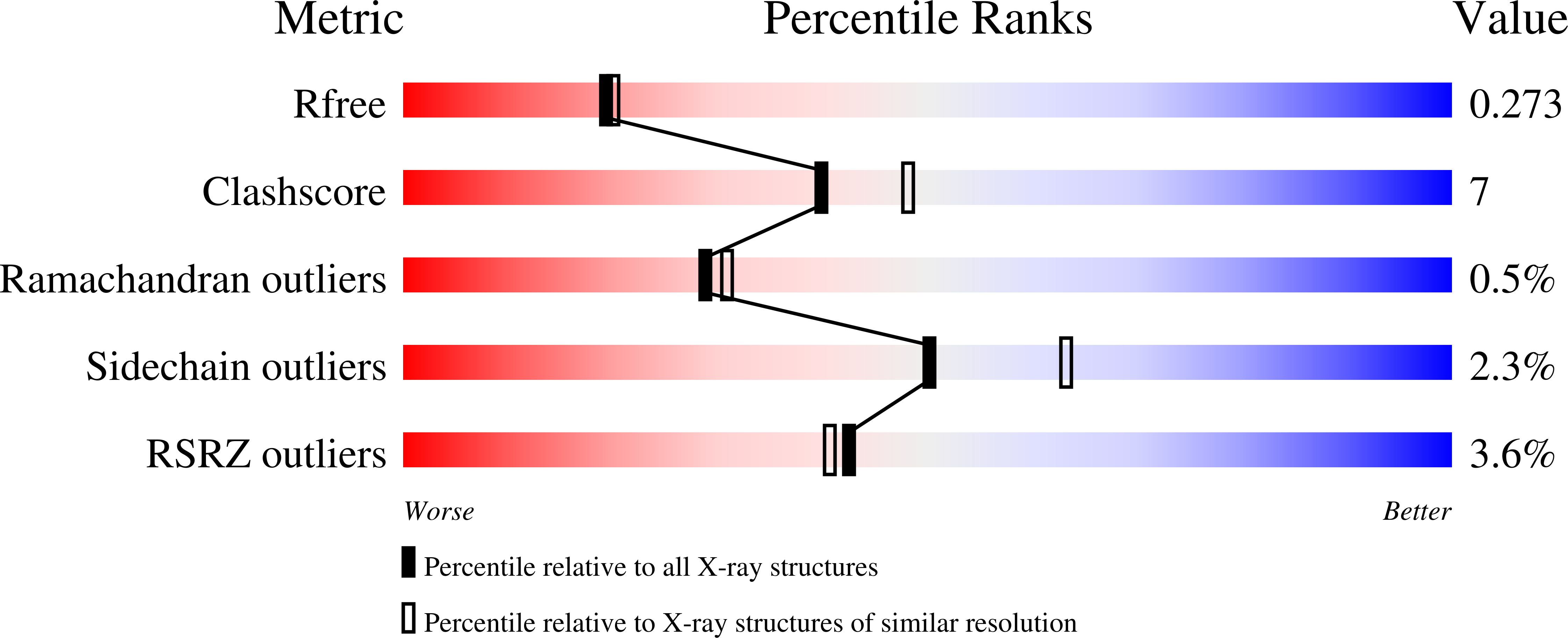
R-Value Free:
0.27
R-Value Work:
0.19
R-Value Observed:
0.20
Space Group:
C 2 2 21