6ZTV image
Entry Detail
PDB ID:
6ZTV
Keywords:
Title:
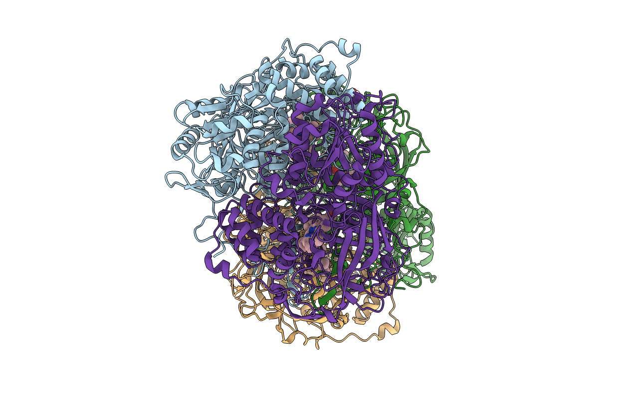
Crystal Structure of catalase HPII from Escherichia coli (serendipitously crystallized)
Biological Source:
Source Organism:
PDB Version:
Deposition Date:
2020-07-20
Release Date:
2020-10-07
Method Details:
Experimental Method:
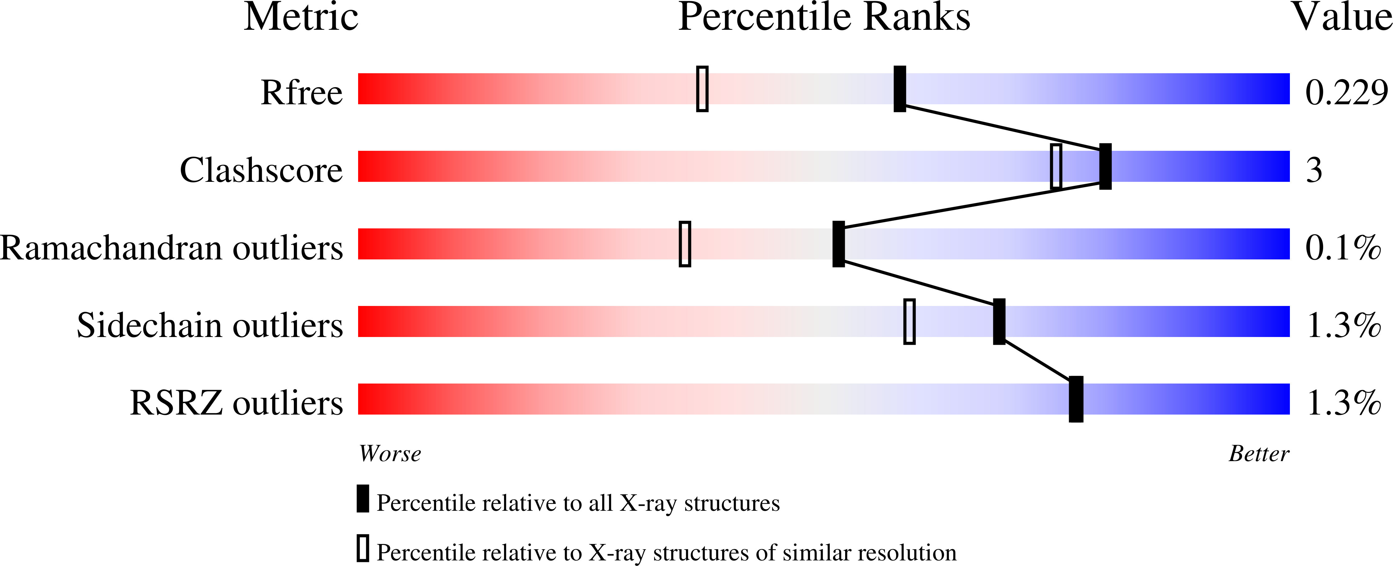
Resolution:
1.78 Å
R-Value Free:
0.22
R-Value Work:
0.18
R-Value Observed:
0.18
Space Group:
P 1 21 1
Macromolecular Entities
Polymer Type:polypeptide(L)
Description:Catalase HPII
Mutations:S99N
Chain IDs:A, B, C, D
Chain Length:753
Number of Molecules:4
Biological Source:Escherichia coli K-12
Modified Residue
Compound ID Chain ID Parent Comp ID Details 2D Image
OCS A CYS modified residue
Primary Citation
Serendipitous crystallization of E. coli HPII catalase, a sequel to "the tale usually not told".
Acta Biochim.Pol. 68 29 31 (2021)
PMID: 33485289 DOI: 10.18388/abp.2020_5501

Abstact

Protein crystallographers are well aware of the trap of crystallizing E. coli proteins instead of the macromolecule of interest if heterologous recombinant protein expression in E. coli was part of the experimental pipeline. Among the well-known culprits are YodA metal-binding lipocalin (25 kDa) and YadF carbonic anhydrase (a tetramer of 25 kDa subunits). We report a novel crystal form of another such culprit, E. coli HPII catalase, which is a tetrameric protein of ~340 kDa molecular weight. HPII is likely to contaminate recombinant protein samples, co-purify, and then co-crystallize with the target proteins, especially if their masses in size exclusion chromatography are ~300-400 kDa. What makes this case more interesting but also parlous, is the fact that HPII can crystallize from very low concentrations, even well below 1 mg/mL.

Legend

Protein

Chemical

Disease

Primary Citation of related structures