
Deposition Date
2020-07-20
Release Date
2020-09-16
Last Version Date
2025-04-02
Entry Detail
PDB ID:
6ZTS
Keywords:
Title:
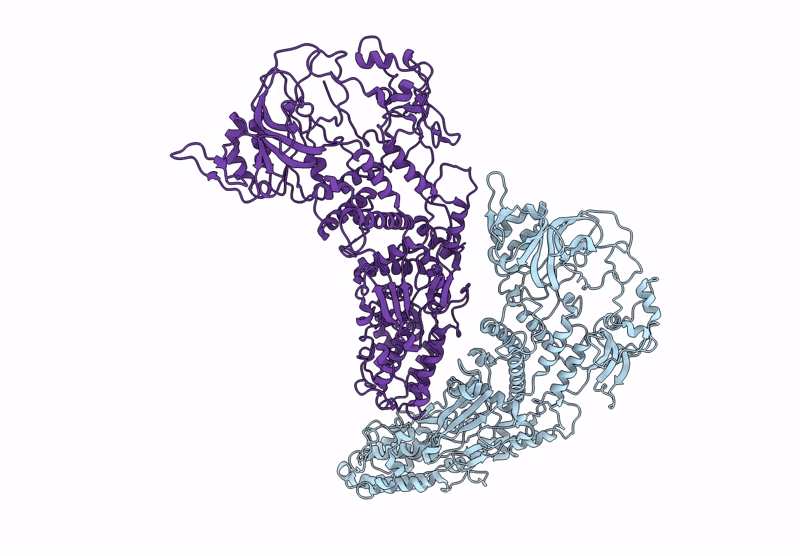
Assembly intermediates of orthoreovirus captured in the cell
Biological Source:
Source Organism(s):
Mammalian orthoreovirus (Taxon ID: 351073)
Method Details:
Experimental Method:
Resolution:
6.60 Å
Aggregation State:
CELL
Reconstruction Method:
TOMOGRAPHY
