6ZQP image
Deposition Date 2020-07-10
Release Date 2020-12-23
Last Version Date 2024-10-23
Entry Detail
PDB ID:
6ZQP
Title:
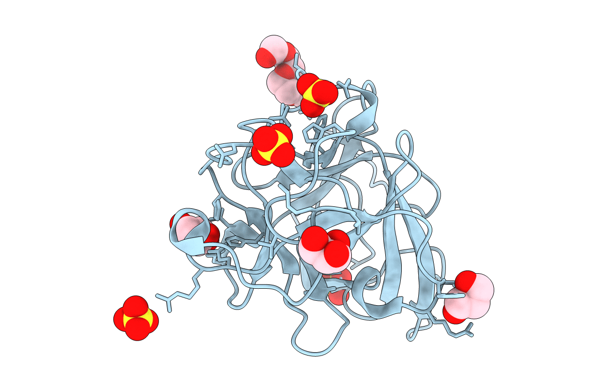
Structure of the Pmt2-MIR domain with bound ligands
Biological Source:
Source Organism:
Host Organism:
Method Details:
Experimental Method:
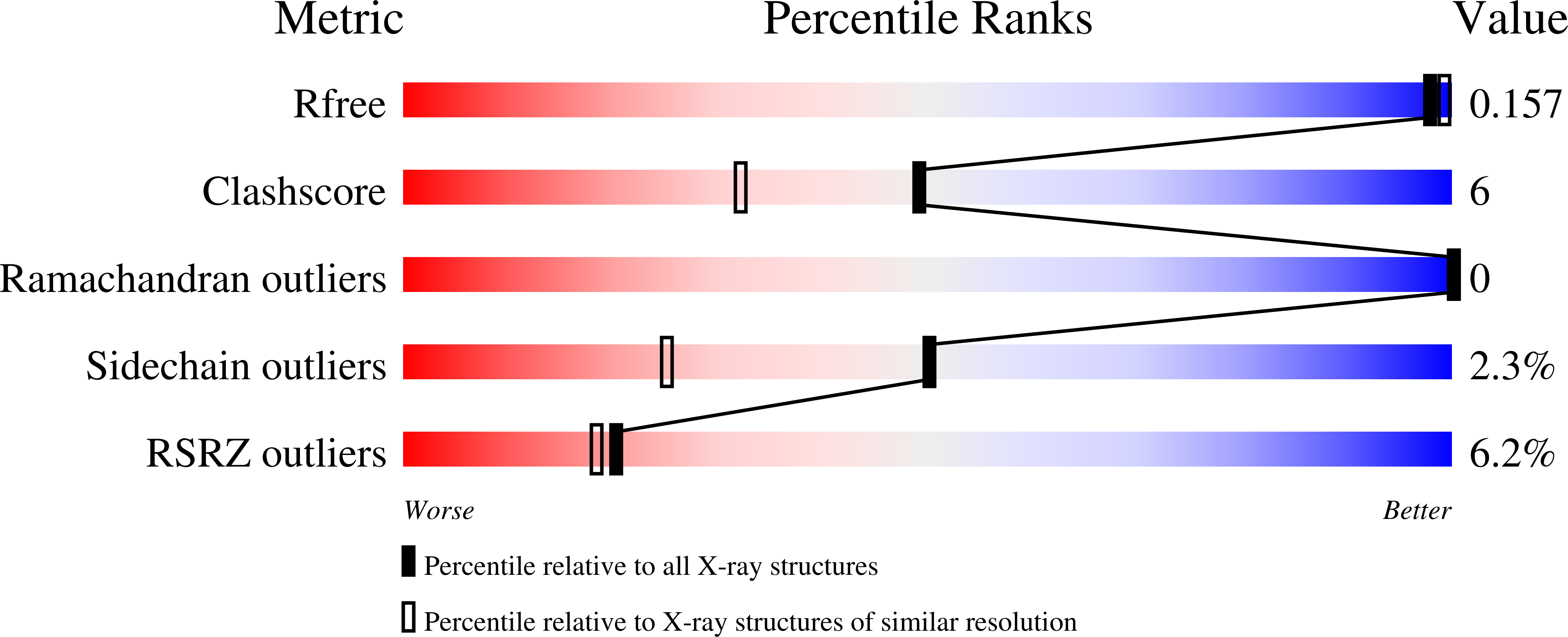
Resolution:
1.60 Å
R-Value Free:
0.15
R-Value Work:
0.14
R-Value Observed:
0.14
Space Group:
P 41 3 2
Macromolecular Entities
Polymer Type:polypeptide(L)
Molecule:PMT2 isoform 1
Chain IDs:A
Chain Length:214
Number of Molecules:1
Biological Source:Saccharomyces cerevisiae
Primary Citation
Functional implications of MIR domains in protein O -mannosylation.
Elife 9 ? ? (2020)
PMID: 33357379 DOI: 10.7554/eLife.61189

Abstact

Protein O-mannosyltransferases (PMTs) represent a conserved family of multispanning endoplasmic reticulum membrane proteins involved in glycosylation of S/T-rich protein substrates and unfolded proteins. PMTs work as dimers and contain a luminal MIR domain with a β-trefoil fold, which is susceptive for missense mutations causing α-dystroglycanopathies in humans. Here, we analyze PMT-MIR domains by an integrated structural biology approach using X-ray crystallography and NMR spectroscopy and evaluate their role in PMT function in vivo. We determine Pmt2- and Pmt3-MIR domain structures and identify two conserved mannose-binding sites, which are consistent with general β-trefoil carbohydrate-binding sites (α, β), and also a unique PMT2-subfamily exposed FKR motif. We show that conserved residues in site α influence enzyme processivity of the Pmt1-Pmt2 heterodimer in vivo. Integration of the data into the context of a Pmt1-Pmt2 structure and comparison with homologous β-trefoil - carbohydrate complexes allows for a functional description of MIR domains in protein O-mannosylation.

Legend

Protein

Chemical

Disease

Primary Citation of related structures