Deposition Date
2020-07-08
Release Date
2020-10-21
Last Version Date
2024-06-19
Entry Detail
PDB ID:
6ZPM
Keywords:
Title:
Crystal structure of the unconventional kinetochore protein Trypanosoma cruzi KKT4 coiled coil domain
Biological Source:
Source Organism(s):
Trypanosoma cruzi (Taxon ID: 5693)
Expression System(s):
Method Details:
Experimental Method:
Resolution:
1.90 Å
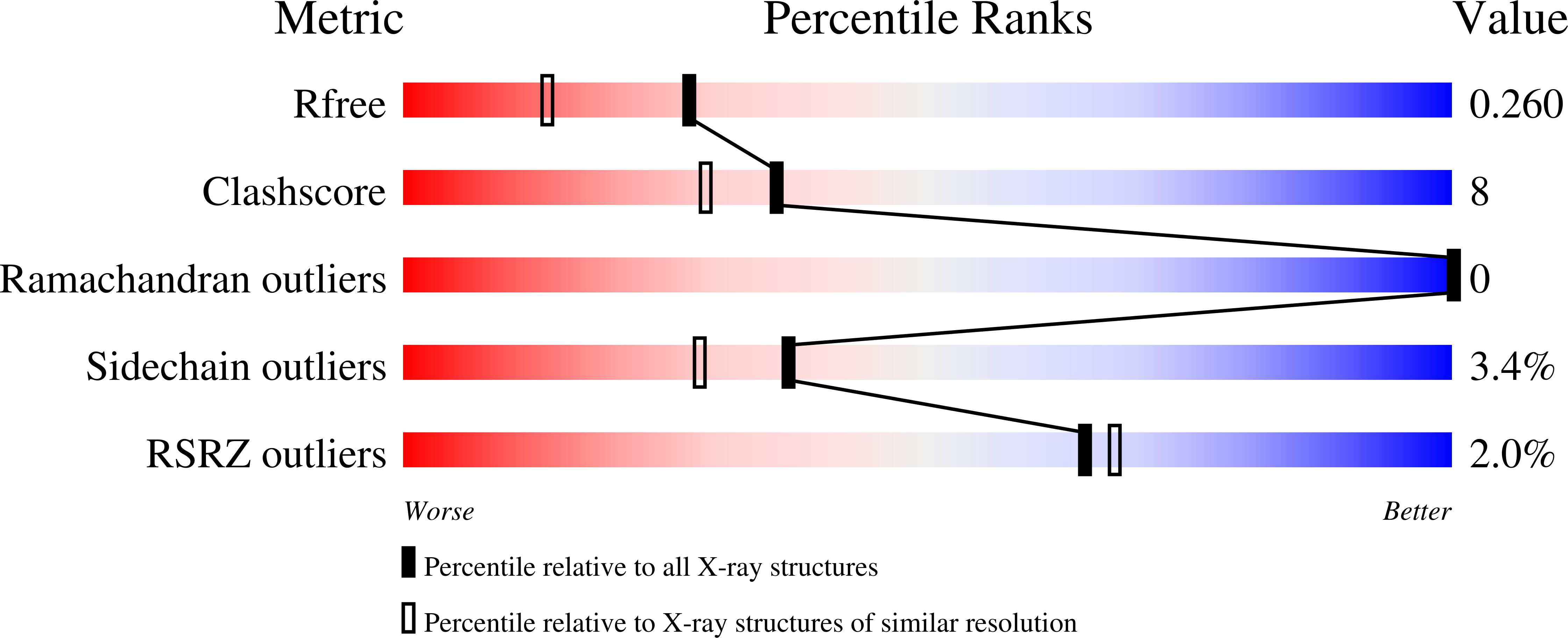
R-Value Free:
0.25
R-Value Work:
0.24
R-Value Observed:
0.24
Space Group:
P 1 21 1