
Deposition Date
2020-07-08
Release Date
2020-12-30
Last Version Date
2025-07-02
Entry Detail
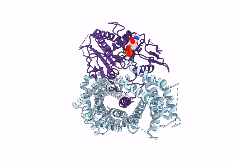
PDB ID:
6ZPH
Keywords:
Title:
Kinesin binding protein complexed with Kif15 motor domain
Biological Source:
Source Organism(s):
Homo sapiens (Taxon ID: 9606)
Expression System(s):
Method Details:
Experimental Method:
Resolution:
6.90 Å
Aggregation State:
PARTICLE
Reconstruction Method:
SINGLE PARTICLE
