6ZMY image
Entry Detail
PDB ID:
6ZMY
Title:
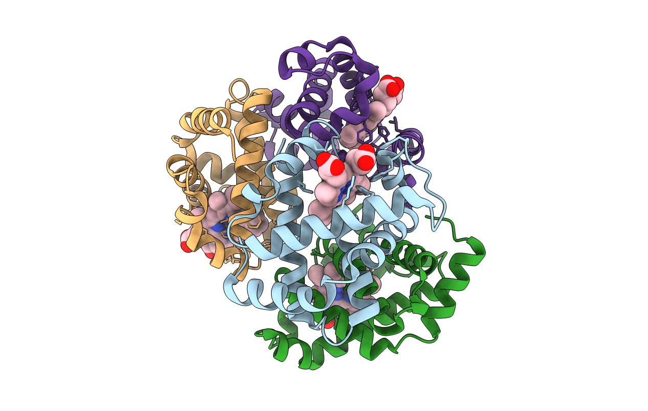
Crystal structure of hemoglobin from turkey (Meleagiris gallopova) crystallized in monoclinic form at 1.66 Angstrom resolution
Biological Source:
Source Organism:
PDB Version:
Deposition Date:
2020-07-04
Release Date:
2020-07-15
Method Details:
Experimental Method:
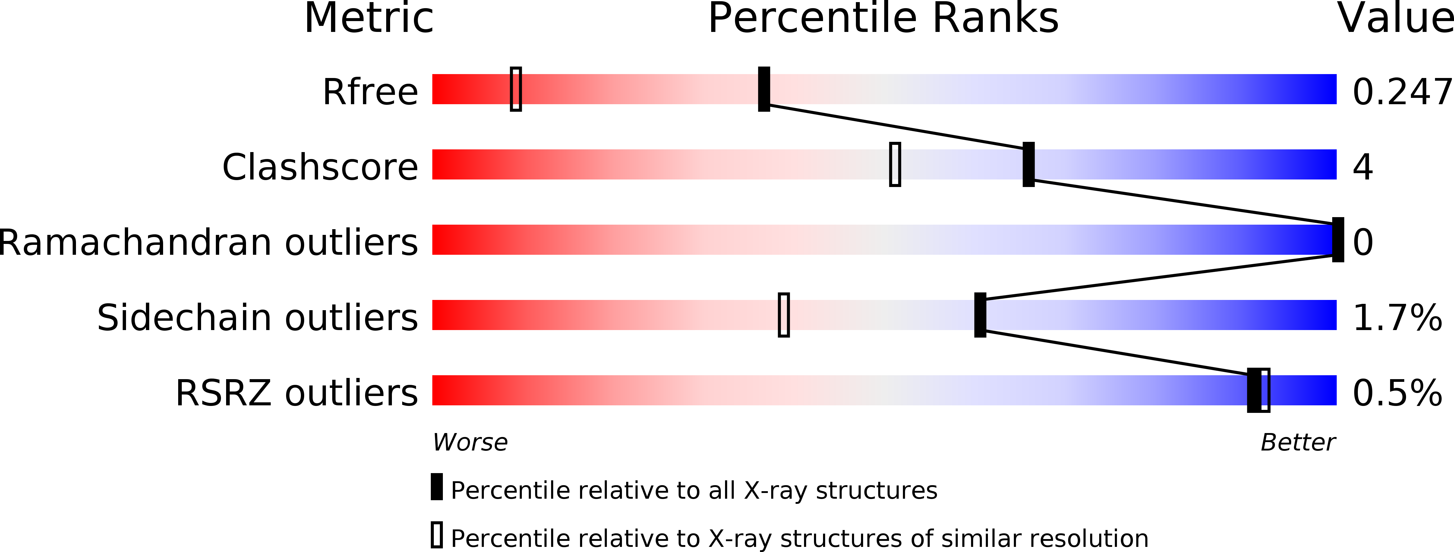
Resolution:
1.66 Å
R-Value Free:
0.25
R-Value Work:
0.20
R-Value Observed:
0.20
Space Group:
P 1 21 1
Macromolecular Entities
Polymer Type:polypeptide(L)
Description:Hemoglobin subunit alpha-A
Chain IDs:A, C
Chain Length:141
Number of Molecules:2
Biological Source:Meleagris gallopavo
Polymer Type:polypeptide(L)
Description:Hemoglobin beta chain
Chain IDs:B, D
Chain Length:146
Number of Molecules:2
Biological Source:Meleagris gallopavo
Ligand Molecules
Primary Citation
Structural studies of hemoglobin from two flightless birds, ostrich and turkey: insights into their differing oxygen-binding properties.
Acta Crystallogr D Struct Biol 77 690 702 (2021)
PMID: 33950023 DOI: 10.1107/S2059798321003417

Abstact

Crystal structures of hemoglobin (Hb) from two flightless birds, ostrich (Struthio camelus) and turkey (Meleagris gallopova), were determined. The ostrich Hb structure was solved to a resolution of 2.22 Å, whereas two forms of turkey Hb were solved to resolutions of 1.66 Å (turkey monoclinic structure; TMS) and 1.39 Å (turkey orthorhombic structure; TOS). Comparison of the amino-acid sequences of ostrich and turkey Hb with those from other avian species revealed no difference in the number of charged residues, but variations were observed in the numbers of hydrophobic and polar residues. Amino-acid-composition-based computation of various physical parameters, in particular their lower inverse transition temperatures and higher average hydrophobicities, indicated that the structures of ostrich and turkey Hb are likely to be highly ordered when compared with other avian Hbs. From the crystal structure analysis, the liganded state of ostrich Hb was confirmed by the presence of an oxygen molecule between the Fe atom and the proximal histidine residue in all four heme regions. In turkey Hb (both TMS and TOS), a water molecule was bound instead of an oxygen molecule in all four heme regions, thus confirming that they assumed the aqua-met form. Analysis of tertiary- and quaternary-structural features led to the conclusion that ostrich oxy Hb and turkey aqua-met Hb adopt the R-/RH-state conformation.

Legend

Protein

Chemical

Disease

Primary Citation of related structures