
Deposition Date
2020-07-02
Release Date
2021-02-24
Last Version Date
2024-05-15
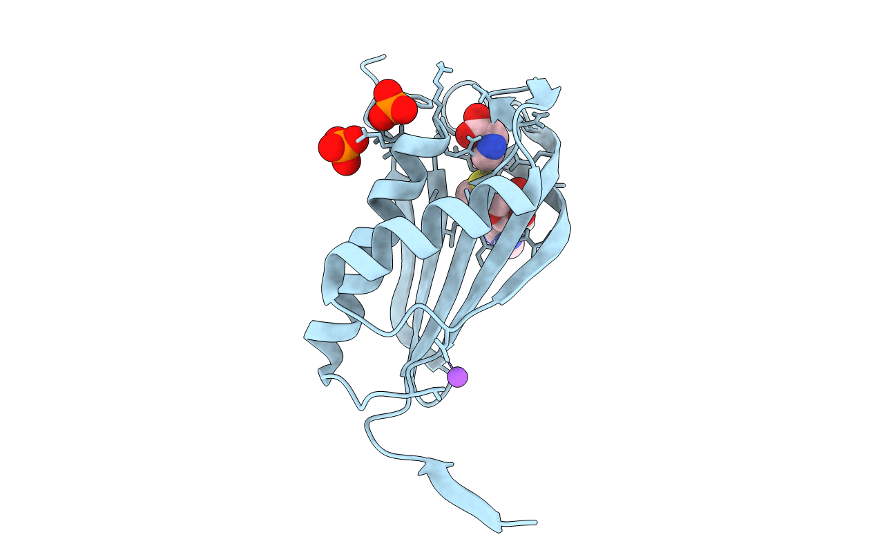
Entry Detail
Biological Source:
Source Organism(s):
unidentified (Taxon ID: 32644)
Expression System(s):
Method Details:
Experimental Method:
Resolution:
1.45 Å
R-Value Free:
0.15
R-Value Work:
0.13
R-Value Observed:
0.13
Space Group:
F 41 3 2
