
Deposition Date
2020-06-30
Release Date
2021-07-07
Last Version Date
2025-07-09
Entry Detail
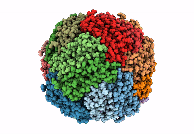
PDB ID:
6ZLG
Keywords:
Title:
Folding of an iron binding peptide in response to sedimentation is resolved using ferritin as a nano-reactor
Biological Source:
Source Organism(s):
Mus musculus (Taxon ID: 10090)
Expression System(s):
Method Details:
Experimental Method:
Resolution:
3.00 Å
Aggregation State:
PARTICLE
Reconstruction Method:
SINGLE PARTICLE
