
Deposition Date
2020-06-24
Release Date
2020-10-14
Last Version Date
2024-05-01
Entry Detail
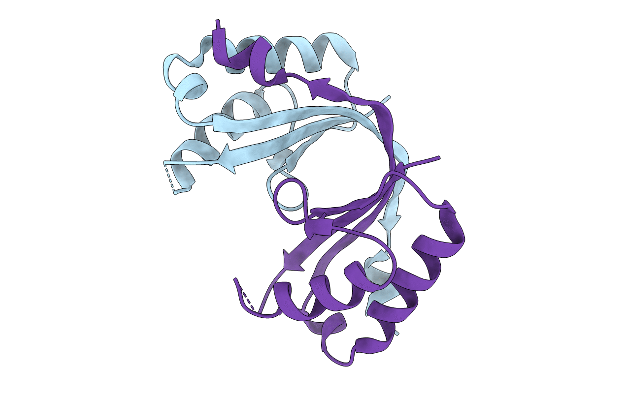
PDB ID:
6ZI1
Keywords:
Title:
Crystal structure of the isolated H. influenzae VapD toxin (D7N mutant)
Biological Source:
Source Organism(s):
Haemophilus influenzae 86-028NP (Taxon ID: 281310)
Expression System(s):
Method Details:
Experimental Method:
Resolution:
2.20 Å
R-Value Free:
0.26
R-Value Work:
0.21
Space Group:
P 3 2 1
