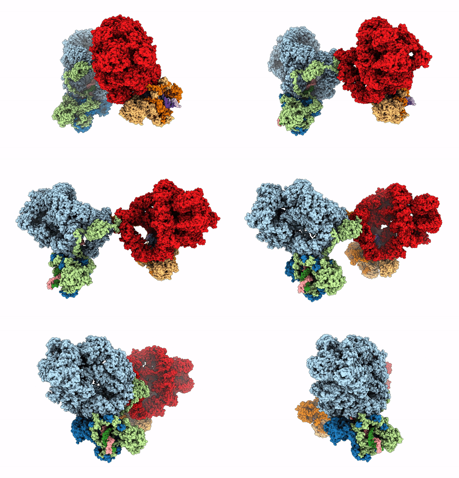
6ZHE image
Deposition Date 2020-06-23
Release Date 2020-10-21
Last Version Date 2024-05-01
Entry Detail
PDB ID:
6ZHE
Title:
Cryo-EM structure of DNA-PK dimer
Biological Source:
Source Organism:
Homo sapiens (Taxon ID: 9606)
Method Details:
Experimental Method:
Resolution:
7.24 Å
Aggregation State:
PARTICLE
Reconstruction Method:
SINGLE PARTICLE
Macromolecular Entities
Structures with similar UniProt ID
Protein Blast
Polymer Type:polypeptide(L)
Molecule:DNA-dependent protein kinase catalytic subunit,DNA-PKcs
Gene (Uniprot):PRKDC
Chain IDs:A, D (auth: F)
Chain Length:4156
Number of Molecules:2
Biological Source:Homo sapiens
Structures with similar UniProt ID
Protein Blast
Polymer Type:polypeptide(L)
Molecule:X-ray repair cross-complementing protein 6
Gene (Uniprot):XRCC6
Chain IDs:B, E (auth: G)
Chain Length:609
Number of Molecules:2
Biological Source:Homo sapiens
Structures with similar UniProt ID
Protein Blast
Polymer Type:polypeptide(L)
Molecule:X-ray repair cross-complementing protein 5
Gene (Uniprot):XRCC5
Chain IDs:C, F (auth: H)
Chain Length:732
Number of Molecules:2
Biological Source:Homo sapiens
Polymer Type:polydeoxyribonucleotide
Molecule:DNA (26-MER)
Chain IDs:I (auth: D)
Chain Length:26
Number of Molecules:1
Biological Source:Homo sapiens
Polymer Type:polydeoxyribonucleotide
Molecule:DNA (28-MER)
Chain IDs:J (auth: E)
Chain Length:28
Number of Molecules:1
Biological Source:Homo sapiens
Polymer Type:polydeoxyribonucleotide
Molecule:DNA (27-MER)
Chain IDs:H (auth: I)
Chain Length:27
Number of Molecules:1
Biological Source:Homo sapiens
Polymer Type:polydeoxyribonucleotide
Molecule:DNA (25-MER)
Chain IDs:G (auth: J)
Chain Length:25
Number of Molecules:1
Biological Source:Homo sapiens
Ligand Molecules
Primary Citation

Abstact

DNA double-strand breaks are the most dangerous type of DNA damage and, if not repaired correctly, can lead to cancer. In humans, Ku70/80 recognizes DNA broken ends and recruits the DNA-dependent protein kinase catalytic subunit (DNA-PKcs) to form DNA-dependent protein kinase holoenzyme (DNA-PK) in the process of non-homologous end joining (NHEJ). We present a 2.8-Å-resolution cryo-EM structure of DNA-PKcs, allowing precise amino acid sequence registration in regions uninterpreted in previous 4.3-Å X-ray maps. We also report a cryo-EM structure of DNA-PK at 3.5-Å resolution and reveal a dimer mediated by the Ku80 C terminus. Central to dimer formation is a domain swap of the conserved C-terminal helix of Ku80. Our results suggest a new mechanism for NHEJ utilizing a DNA-PK dimer to bring broken DNA ends together. Furthermore, drug inhibition of NHEJ in combination with chemo- and radiotherapy has proved successful, making these models central to structure-based drug targeting efforts.

Legend

Protein

Chemical

Disease

Primary Citation of related structures