6ZCO image
Entry Detail
PDB ID:
6ZCO
Keywords:
Title:
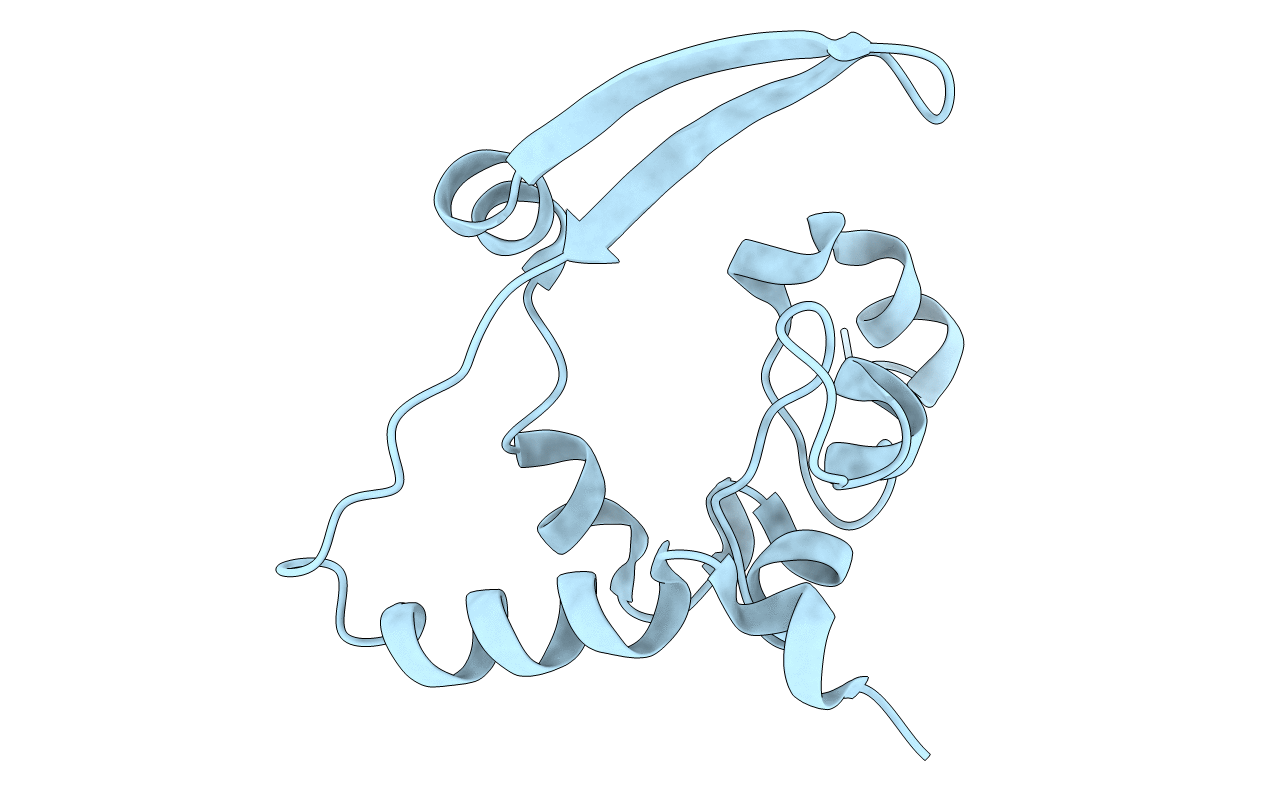
Crystal Structure of C-terminal Dimerization Domain of Nucleocapsid Phosphoprotein from SARS-CoV-2, crystal form II
Biological Source:
PDB Version:
Deposition Date:
2020-06-11
Release Date:
2020-07-01
Method Details:
Experimental Method:
Resolution:
1.36 Å
R-Value Free:
0.19
R-Value Work:
0.14
R-Value Observed:
0.15
Space Group:
I 41
Macromolecular Entities
Polymer Type:polypeptide(L)
Description:Nucleoprotein
Chain IDs:A
Chain Length:135
Number of Molecules:1
Biological Source:Severe acute respiratory syndrome coronavirus 2
Primary Citation
High-resolution structure and biophysical characterization of the nucleocapsid phosphoprotein dimerization domain from the Covid-19 severe acute respiratory syndrome coronavirus 2.
Biochem.Biophys.Res.Commun. 538 54 62 (2021)
PMID: 33039147 DOI: 10.1016/j.bbrc.2020.09.131

Abstact

Unprecedented by number of casualties and socio-economic burden occurring worldwide, the coronavirus disease 2019 (Covid-19) pandemic caused by the severe acute respiratory syndrome coronavirus 2 (SARS-CoV-2) is the worst health crisis of this century. In order to develop adequate countermeasures against Covid-19, identification and structural characterization of suitable antiviral targets within the SARS-CoV-2 protein repertoire is urgently needed. The nucleocapsid phosphoprotein (N) is a multifunctional and highly immunogenic determinant of virulence and pathogenicity, whose main functions consist in oligomerizing and packaging the single-stranded RNA (ssRNA) viral genome. Here we report the structural and biophysical characterization of the SARS-CoV-2 N C-terminal domain (CTD), on which both N homo-oligomerization and ssRNA binding depend. Crystal structures solved at 1.44 Å and 1.36 Å resolution describe a rhombus-shape N CTD dimer, which stably exists in solution as validated by size-exclusion chromatography coupled to multi-angle light scattering and analytical ultracentrifugation. Differential scanning fluorimetry revealed moderate thermal stability and a tendency towards conformational change. Microscale thermophoresis demonstrated binding to a 7-bp SARS-CoV-2 genomic ssRNA fragment at micromolar affinity. Furthermore, a low-resolution preliminary model of the full-length SARS-CoV N in complex with ssRNA, obtained by cryo-electron microscopy, provides an initial understanding of self-associating and RNA binding functions exerted by the SARS-CoV-2 N.

Legend

Protein

Chemical

Disease

Primary Citation of related structures