6Z7P image
Deposition Date 2020-06-01
Release Date 2020-07-15
Last Version Date 2024-05-15
Entry Detail
PDB ID:
6Z7P
Title:
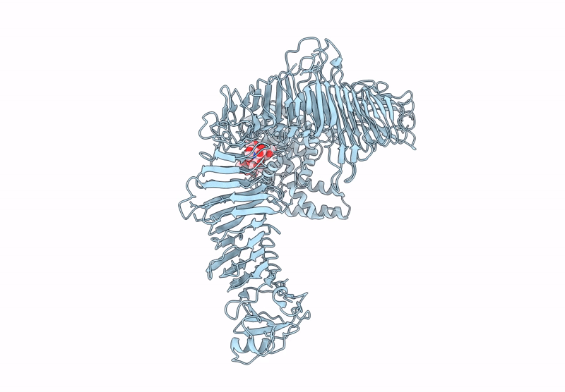
Composite model of the Caulobacter crescentus S-layer bound to the O-antigen of lipopolysaccharide
Biological Source:
Method Details:
Experimental Method:
Resolution:
4.80 Å
Aggregation State:
CELL
Reconstruction Method:
SUBTOMOGRAM AVERAGING
Macromolecular Entities
Structures with similar UniProt ID
Protein Blast
Polymer Type:polypeptide(L)
Molecule:S-layer protein
Gene (Uniprot):rsaA
Chain IDs:A
Chain Length:1025
Number of Molecules:1
Biological Source:Caulobacter vibrioides (strain ATCC 19089 / CB15)
Ligand Molecules
Primary Citation

Abstact

Most bacterial and all archaeal cells are encapsulated by a paracrystalline, protective, and cell-shape-determining proteinaceous surface layer (S-layer). On Gram-negative bacteria, S-layers are anchored to cells via lipopolysaccharide. Here, we report an electron cryomicroscopy structure of the Caulobacter crescentus S-layer bound to the O-antigen of lipopolysaccharide. Using native mass spectrometry and molecular dynamics simulations, we deduce the length of the O-antigen on cells and show how lipopolysaccharide binding and S-layer assembly is regulated by calcium. Finally, we present a near-atomic resolution in situ structure of the complete S-layer using cellular electron cryotomography, showing S-layer arrangement at the tip of the O-antigen. A complete atomic structure of the S-layer shows the power of cellular tomography for in situ structural biology and sheds light on a very abundant class of self-assembling molecules with important roles in prokaryotic physiology with marked potential for synthetic biology and surface-display applications.

Legend

Protein

Chemical

Disease

Primary Citation of related structures