Deposition Date
2020-05-22
Release Date
2020-12-09
Last Version Date
2024-05-22
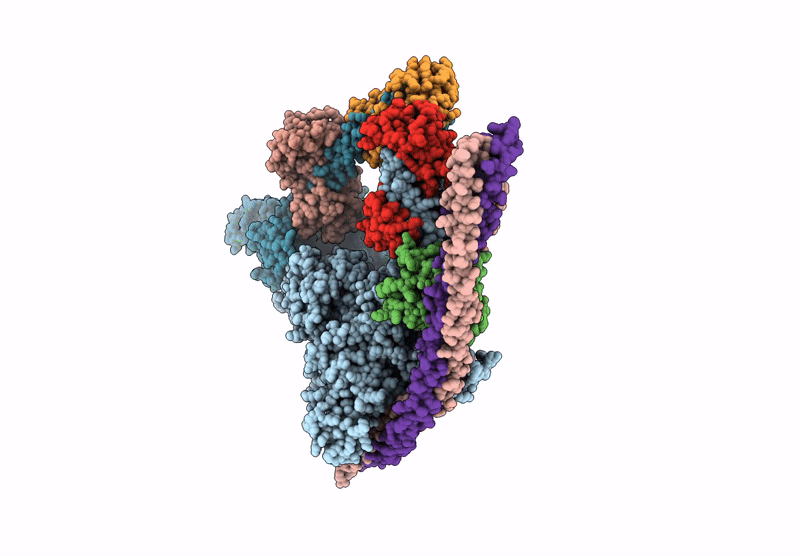
Method Details:
Experimental Method:
Resolution:
6.30 Å
Aggregation State:
PARTICLE
Reconstruction Method:
SINGLE PARTICLE