6Z3U image
Deposition Date 2020-05-22
Release Date 2020-12-09
Last Version Date 2024-01-24
Entry Detail
PDB ID:
6Z3U
Keywords:
Title:
Structure of the CAK complex form Chaetomium thermophilum
Biological Source:
Method Details:
Experimental Method:
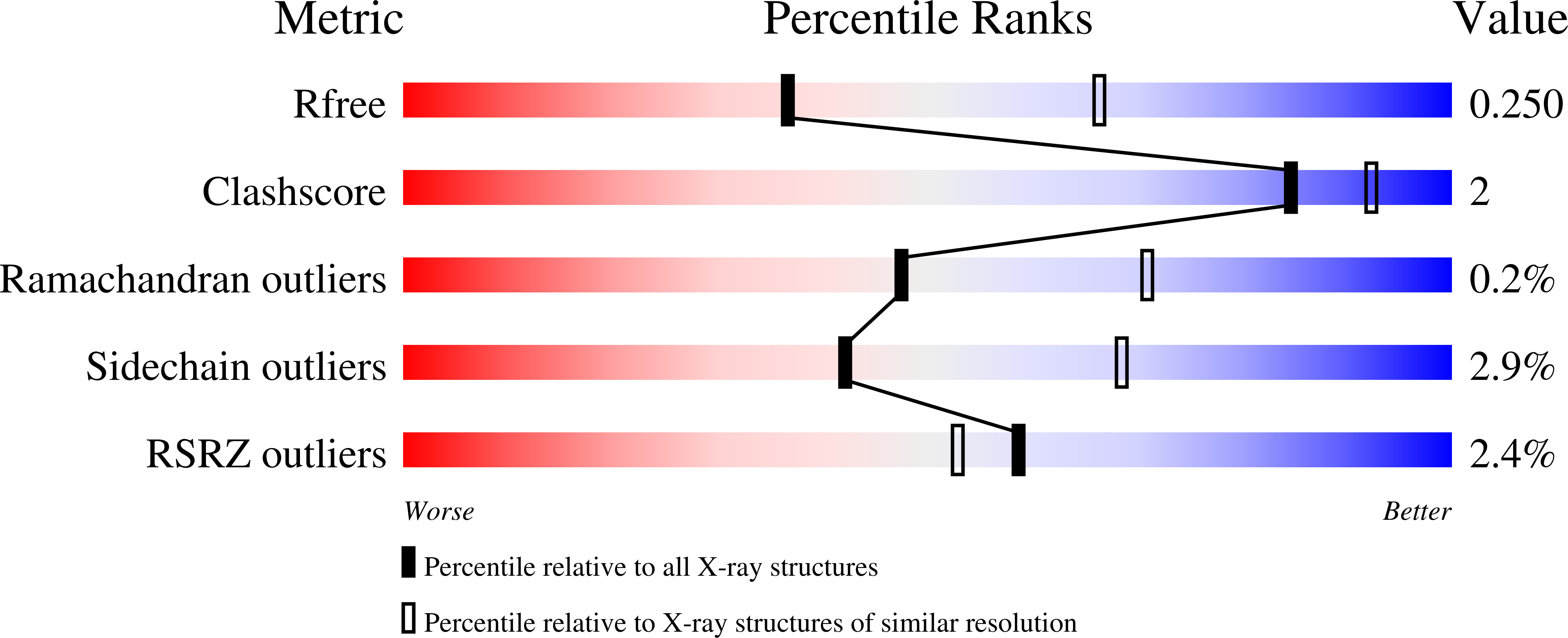
Resolution:
2.60 Å
R-Value Free:
0.24
R-Value Work:
0.20
R-Value Observed:
0.20
Space Group:
P 1 21 1
Macromolecular Entities
Polymer Type:polypeptide(L)
Molecule:CYCLIN domain-containing protein
Gene (Uniprot):CTHT_0069020
Chain IDs:A, D
Chain Length:425
Number of Molecules:2
Biological Source:Chaetomium thermophilum (strain DSM 1495 / CBS 144.50 / IMI 039719)
Polymer Type:polypeptide(L)
Molecule:Protein kinase domain-containing protein
Gene (Uniprot):CTHT_0061570
Chain IDs:B, E
Chain Length:437
Number of Molecules:2
Biological Source:Chaetomium thermophilum (strain DSM 1495 / CBS 144.50 / IMI 039719)
Polymer Type:polypeptide(L)
Molecule:RING-type domain-containing protein
Gene (Uniprot):CTHT_0060790
Chain IDs:C, F
Chain Length:69
Number of Molecules:2
Biological Source:Chaetomium thermophilum (strain DSM 1495 / CBS 144.50 / IMI 039719)
Ligand Molecules
Primary Citation
Structural basis for CDK7 activation by MAT1 and Cyclin H.
Proc.Natl.Acad.Sci.USA 117 26739 26748 (2020)
PMID: 33055219 DOI: 10.1073/pnas.2010885117

Abstact

Cyclin-dependent kinase 7 (CDK7), Cyclin H, and the RING-finger protein MAT1 form the heterotrimeric CDK-activating kinase (CAK) complex which is vital for transcription and cell-cycle control. When associated with the general transcription factor II H (TFIIH) it activates RNA polymerase II by hyperphosphorylation of its C-terminal domain (CTD). In the absence of TFIIH the trimeric complex phosphorylates the T-loop of CDKs that control cell-cycle progression. CAK holds a special position among the CDK branch due to this dual activity and the dependence on two proteins for activation. We solved the structure of the CAK complex from the model organism Chaetomium thermophilum at 2.6-Å resolution. Our structure reveals an intricate network of interactions between CDK7 and its two binding partners MAT1 and Cyclin H, providing a structural basis for the mechanism of CDK7 activation and CAK activity regulation. In vitro activity measurements and functional mutagenesis show that CDK7 activation can occur independent of T-loop phosphorylation and is thus exclusively MAT1-dependent by positioning the CDK7 T-loop in its active conformation.

Legend

Protein

Chemical

Disease

Primary Citation of related structures